Table of contents:
Introduction to SUV Pet Scans

Single-photon emission computed tomography (SPECT) and positron emission tomography (PET) scans are crucial diagnostic tools in modern medicine. SUV pet scans, a specific type of PET scan, provide valuable information about metabolic activity within tissues, aiding in the detection and characterization of various medical conditions. These scans are particularly useful in oncology, where they assist in tumor staging, treatment response monitoring, and recurrence detection.
SUV pet scans leverage the principles of radioactivity to visualize metabolic processes. A radiotracer, a substance containing a radioactive isotope, is administered to the patient. The radiotracer accumulates in areas of higher metabolic activity, which are then visualized using specialized imaging equipment. This allows clinicians to identify areas of increased metabolic activity, often indicative of abnormal cellular growth or other pathological processes.
Radiotracer Principles
Radiotracers are crucial to SUV pet scans, as they provide the means to visualize metabolic activity. These molecules contain a radioactive isotope that emits positrons, which, upon colliding with electrons, produce gamma rays. These gamma rays are detected by the PET scanner, allowing the creation of a 3D image reflecting the distribution of the radiotracer within the body. The radiotracer’s choice is critical, as different radiotracers target specific metabolic pathways, thus influencing the image’s interpretation.
Types of SUV Pet Scans
While the core principle remains consistent, different radiotracers and imaging protocols can be used, yielding varying types of SUV PET scans. These differences primarily relate to the specific metabolic pathways targeted by the radiotracer. For instance, a scan focused on glucose metabolism will provide a different image profile compared to one focusing on amino acid metabolism.
Common Radiotracers and Their Applications
| Radiotracer | Application | Description |
|---|---|---|
| Fluorodeoxyglucose (FDG) | Oncology, neurology, and cardiology | The most commonly used radiotracer, FDG is a glucose analog that accumulates in tissues with high metabolic activity, particularly cancerous tumors. |
| 18F-Choline | Oncology | This radiotracer is concentrated in areas of rapid cell division, making it useful in detecting and characterizing certain types of tumors. |
| 18F-Dopamine | Neurology | This radiotracer can be used to evaluate dopamine receptors in the brain, aiding in the diagnosis of neurological disorders like Parkinson’s disease. |
| 18F-Fluorothymidine (FLT) | Oncology | FLT targets rapidly dividing cells, providing information on tumor proliferation and response to treatment. |
| 11C-Methionine | Oncology | This radiotracer focuses on amino acid metabolism, aiding in the detection and characterization of certain tumors. |
SUV Pet Scan Indications and Applications
SUV PET scans, or standardized uptake values Positron Emission Tomography scans, provide crucial insights into metabolic activity within the body. This information is particularly valuable in identifying and characterizing various medical conditions, especially those involving cellular processes, such as cancer. By visualizing areas of high metabolic activity, SUV PET scans can aid in diagnosis, treatment planning, and monitoring.
The ability to assess tumor activity and response to therapy is a key application of SUV PET scans. These scans provide a unique perspective on the biological behavior of tumors, supplementing the information obtained from anatomical imaging techniques like CT and MRI. This multifaceted approach allows for a more comprehensive understanding of the disease process and facilitates more effective treatment strategies.
Medical Conditions Where SUV Pet Scans Are Commonly Used
SUV PET scans are frequently employed in the diagnosis and management of various medical conditions. They are particularly useful in oncology, but also find applications in neurology and cardiology. Their ability to visualize metabolic activity sets them apart from other imaging modalities.
- Oncology: SUV PET scans are highly valuable in detecting primary tumors, staging cancer, assessing response to therapy, and detecting recurrent disease. They can identify areas of high metabolic activity, often indicative of malignancy, allowing for more precise localization and characterization of tumors. For instance, a patient suspected of having lung cancer could benefit from an SUV PET scan to determine the extent of the disease and guide treatment decisions.
- Neurology: SUV PET scans can assist in the diagnosis of neurodegenerative diseases like Alzheimer’s disease and Parkinson’s disease by highlighting areas of altered metabolic activity in the brain. This can provide crucial information for evaluating the progression of the disease and potential treatment response.
- Cardiology: SUV PET scans can assess myocardial viability and metabolic function in patients with suspected coronary artery disease. By measuring the metabolic activity of the heart muscle, these scans can help determine the extent of damage and guide treatment decisions.
Clinical Situations Where SUV Pet Scans Are Valuable
SUV PET scans are a valuable diagnostic tool in various clinical situations. Their unique ability to visualize metabolic activity provides a crucial perspective in evaluating the extent and nature of disease.
- Suspected Cancer Recurrence: In patients with a history of cancer, SUV PET scans can identify areas of abnormal metabolic activity that might indicate a recurrence, allowing for prompt intervention and treatment.
- Staging of Cancer: SUV PET scans can help determine the extent of cancer spread, differentiating between localized and metastatic disease. This crucial information allows for appropriate staging and subsequent treatment strategies.
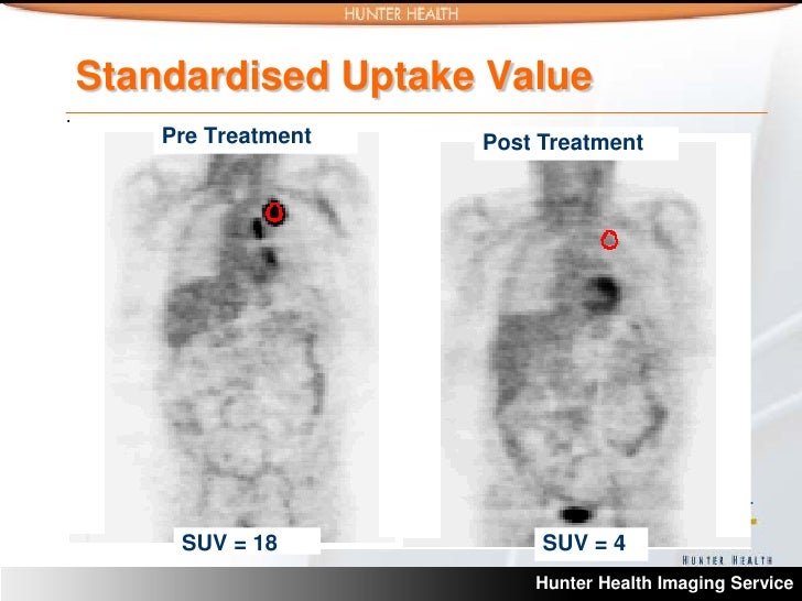
- Response to Therapy Monitoring: SUV PET scans can assess the effectiveness of treatment by monitoring the metabolic activity of the tumor over time. A reduction in SUV values can indicate a positive response to treatment, while an increase may signal resistance or recurrence.
SUV Pet Scans in Cancer Diagnosis and Treatment Monitoring
SUV PET scans play a pivotal role in cancer diagnosis and treatment monitoring. Their ability to visualize metabolic activity provides valuable insights into tumor behavior and response to therapy.
- Tumor Activity Assessment: SUV PET scans provide a quantitative measure of tumor metabolic activity. High SUV values typically correlate with higher tumor activity, while lower values suggest reduced activity. This allows clinicians to assess the aggressiveness of the tumor and monitor its response to treatment.
- Response to Therapy Evaluation: By tracking SUV values over time, clinicians can assess the effectiveness of treatment. A decrease in SUV values indicates that the therapy is impacting the tumor’s metabolic activity, suggesting a positive response. Conversely, an increase in SUV values could signal treatment resistance or recurrence.
Comparison of SUV Pet Scan Findings with Other Imaging Techniques
The following table compares SUV PET scan findings with those of CT and MRI scans.
| Imaging Technique | SUV Pet Scan Findings | CT Scan Findings | MRI Findings |
|---|---|---|---|
| SUV PET Scan | Metabolic activity (e.g., high SUV values in areas of high metabolic activity, often indicative of malignancy); Provides functional information about the tissue | Anatomical details (e.g., tumor size, shape, location); Provides anatomical information about the tissue | Soft tissue contrast (e.g., edema, inflammation); Provides high-resolution soft tissue images |
SUV Pet Scan Interpretation and Reporting
Interpreting SUV (standardized uptake value) PET scan results requires careful consideration of various factors influencing the values. Accurate interpretation is crucial for clinical decision-making, guiding treatment strategies, and ultimately, improving patient outcomes. This section delves into the nuances of SUV value analysis, reporting procedures, and the integration of findings into the overall patient picture.
SUV values are not absolute measurements but rather relative indicators of metabolic activity within a specific region of interest. Understanding the factors that influence these values, including patient preparation, scanner settings, and physiological variations, is essential for a thorough and accurate interpretation.
Factors Influencing SUV Values
Various factors can affect SUV values, necessitating careful consideration during interpretation. Patient factors, such as hydration status, recent meals, and concurrent medications, can significantly impact metabolic activity and, consequently, SUV values. Scanner settings, including the specific PET scanner model, calibration protocols, and acquisition parameters, also influence the precision of SUV measurements. Finally, the physiological variations between patients, including differences in age, body composition, and underlying disease processes, need to be factored into the analysis.
Methods for Interpreting SUV Values
SUV values are typically interpreted by comparing them to normal values or standard uptake values (SUVs). Normal values are established through large-scale studies involving healthy individuals, providing a baseline for comparison. Standard uptake values (SUVmax, SUVmean, SUVpeak) represent the maximal, average, and peak metabolic activity in specific anatomical locations. By comparing the patient’s SUV values to these reference points, clinicians can assess the relative metabolic activity of a given region.
SUV Pet Scan Reporting Procedures
Reporting SUV PET scan results involves a structured approach encompassing image analysis and data interpretation. Radiologists meticulously evaluate the images for any abnormalities, paying close attention to the location, size, and shape of the lesions. Quantitative analysis of SUV values, including calculations of SUVmax and SUVmean, provides further insight into the metabolic activity of identified regions. The reporting process also involves noting any clinical correlations and integrating them with the patient’s history, physical examination findings, and other diagnostic tests.
Integration of SUV Findings into the Patient’s Clinical Picture
The interpretation of SUV PET scan findings is not isolated but rather integrated into the broader clinical context. Radiologists consider the patient’s medical history, symptoms, physical examination, and results from other imaging modalities. For example, a high SUV value in a known tumor region can support the diagnosis and guide treatment decisions. Conversely, a low SUV value in a suspected area of disease may warrant further investigation. The integration of these findings into the overall clinical picture allows for a more comprehensive understanding of the patient’s condition and facilitates informed decision-making.
Examples of SUV PET Scan Reports and Key Findings
A typical SUV PET scan report may include a description of the scan protocol, the identified anatomical locations, and the calculated SUV values. The report will likely detail any significant findings, such as increased SUV values in a suspected tumor site, potentially indicating malignancy or inflammation. Conversely, decreased SUV values in a previously identified tumor region might suggest disease response to treatment. An example of a report could highlight an SUVmax value of 10 in a right-lobe lung lesion, significantly higher than the surrounding lung tissue, suggestive of lung cancer. Alternatively, a patient with a known history of lymphoma might exhibit a decrease in SUVmax in the affected lymph nodes following chemotherapy, potentially indicating a positive response to treatment. These are just a few examples, and each report should be carefully evaluated in the context of the individual patient’s clinical presentation.
Limitations and Considerations
SUV PET scans, while powerful tools, are not without limitations. Factors such as patient preparation, physiological variations, and the inherent nature of the technique can influence the accuracy and interpretation of results. Understanding these limitations is crucial for clinicians to avoid misdiagnosis and ensure appropriate patient management.
Factors Affecting Accuracy
Several factors can impact the accuracy of SUV PET scan results. Patient factors, such as hydration status, concurrent medications, and physiological variations, can affect tracer uptake and ultimately impact the calculated SUV values. Furthermore, technical factors, such as scan quality, equipment calibration, and the expertise of the personnel involved, can also contribute to variations in results.
Potential Risks and Side Effects
SUV PET scans, while generally safe, do involve exposure to ionizing radiation. The radiation dose is relatively low compared to some other imaging modalities, but it is important to consider this aspect, particularly for repeated scans or scans in vulnerable populations. The risks are generally minimal, but allergic reactions to the injected radiotracer are a rare but possible complication.
Alternative Diagnostic Modalities
Several alternative diagnostic modalities can complement SUV PET scans in specific clinical scenarios. MRI (Magnetic Resonance Imaging) can provide detailed anatomical information, particularly for assessing soft tissue structures. CT (Computed Tomography) scans are valuable for visualizing bone structures and identifying potential anatomical abnormalities that may be influencing the results. Biopsies can be utilized to obtain tissue samples for further histological analysis, especially in cases of indeterminate findings on imaging studies. These modalities often offer complementary information that enhances the overall diagnostic accuracy.
Pre-Scan Preparation
Proper pre-scan preparation is essential for obtaining reliable SUV PET scan results. Patients are often instructed to fast for a specific period before the scan to minimize the impact of recent food intake on tracer uptake. Hydration status can also influence the results, so patients may be instructed to maintain their usual fluid intake. Concurrent medications can affect tracer uptake and should be reported to the radiologist.
Post-Scan Care
Post-scan care procedures are generally straightforward. Patients are typically monitored for any adverse reactions to the radiotracer. The radiologist will provide specific instructions regarding activity levels and fluid intake post-scan, which may vary depending on the patient’s condition and the specific radiotracer used. It is important to follow these instructions meticulously to ensure optimal recovery and to avoid potential complications.
Evaluating Potential Confounding Factors
Patient demographics, medical history, and concurrent medications can all influence the interpretation of SUV PET scan results. Age, gender, and body mass index can affect tracer uptake, so these factors should be considered when evaluating the findings. A detailed patient history, including prior illnesses, surgeries, and medications, can help to identify potential confounding factors that may impact the results. The presence of concurrent medications, especially those known to influence metabolism or organ function, can significantly influence the results.
Future Trends and Developments
The field of SUV PET scans is experiencing rapid evolution, driven by advancements in radiotracer technology, imaging techniques, and computational analysis. These innovations are poised to significantly enhance the diagnostic capabilities and therapeutic applications of these scans, leading to improved patient outcomes.
Current Research and Development Efforts
Ongoing research focuses on developing more sensitive and specific radiotracers that target particular molecular markers associated with disease. This includes the design of radiopharmaceuticals that bind with higher affinity to specific receptors or proteins involved in cancer growth and progression. Further development in PET scanner technology aims to improve spatial resolution and reduce radiation dose to patients. These advancements will enable clinicians to acquire high-quality images with minimal exposure to ionizing radiation.
Advancements in Radiotracer Development
Novel radiotracers are being developed to provide more detailed information about disease processes. These include radioligands targeting specific receptor subtypes, allowing for more precise characterization of tumor heterogeneity. Fluorescent-based PET tracers are also under investigation, enabling dual-modality imaging with CT or MRI for enhanced visualization and comprehensive analysis. This development enables more accurate delineation of tumor boundaries and their surrounding microenvironment, improving surgical planning and radiation therapy targeting.
Advancements in Imaging Techniques
Innovations in PET imaging techniques are enhancing image quality and reducing acquisition time. The use of iterative reconstruction algorithms improves image resolution and signal-to-noise ratio, leading to a clearer visualization of metabolic activity within the tissues. Hybrid PET/MRI scanners combine the advantages of both modalities, providing anatomical and functional information in a single scan. This integration offers a comprehensive view of the tumor and surrounding tissue, allowing for a more precise assessment of tumor extent and characteristics.
Emerging Applications in Personalized Medicine and Targeted Therapies
SUV PET scans are increasingly employed in personalized medicine to tailor treatment strategies based on individual patient characteristics. The ability to assess metabolic activity and tumor heterogeneity enables clinicians to select therapies that are most likely to be effective for a particular patient. This approach will lead to more effective and less toxic therapies. The integration of SUV PET scan data with genomic profiling further refines patient stratification, allowing for targeted therapies based on the specific genetic makeup of the tumor.
Expected Evolution of SUV PET Scan Technology
Future SUV PET scans are expected to become more integrated into the clinical workflow, facilitating real-time decision-making. This integration will likely involve the use of artificial intelligence (AI) and machine learning (ML) algorithms to analyze the vast amount of data generated by PET scans. AI-powered tools will assist in identifying subtle patterns and anomalies, enabling earlier detection and more accurate diagnosis of various diseases. The increased use of AI will also assist in the interpretation and reporting of PET scans, leading to improved diagnostic accuracy and reduced diagnostic delays. Ultimately, this will lead to more personalized and effective treatments for a wider range of conditions.