Table of contents:
- Introduction to SUV 5.2 PET Scan
- Different Types of SUVs in PET Scans
- Clinical Applications of SUV 5.2 PET Scan
- Interpretation and Reporting of SUV 5.2 PET Scan Results
- Limitations and Potential Pitfalls of SUV 5.2 PET Scan
- Comparison with Other Imaging Modalities
- Future Directions and Advancements in SUV PET Scan Technology
Introduction to SUV 5.2 PET Scan
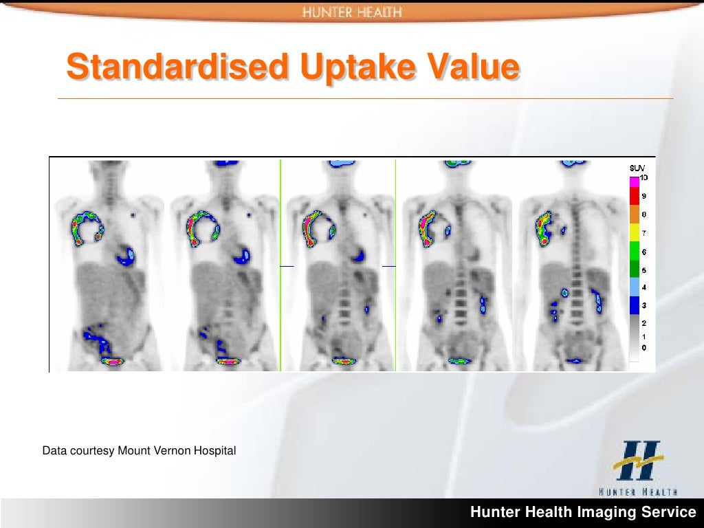
Positron emission tomography (PET) scans are powerful diagnostic tools that visualize metabolic activity within the body. A 5.2 SUV PET scan is a specialized type of PET scan that measures the standardized uptake value (SUV) of a radiotracer, typically glucose, in various tissues. This measurement helps physicians identify areas of increased metabolic activity, which can be indicative of certain diseases, particularly cancers.
The 5.2 SUV value is a standardized way to quantify the concentration of the radiotracer in a particular region of the body relative to a reference standard. A higher SUV value suggests a higher uptake of the radiotracer, which could indicate an area of abnormal metabolic activity, potentially signifying the presence of cancer or other pathological processes. The 5.2 designation refers to a specific range or threshold used for interpretation.
SUV Measurement in PET Scans
The standardized uptake value (SUV) is a crucial metric in interpreting PET scan results. It is calculated by dividing the metabolic activity in a region of interest by the activity in a reference region, typically the liver or blood. This normalization allows for comparisons across different patients and scans. The SUV value is often presented as a numerical value (e.g., SUV 5.2), and it helps clinicians understand the degree of metabolic activity within specific regions. This standardized approach ensures consistent and accurate interpretation of the scan results.
Patient Preparation for a 5.2 SUV PET Scan
Proper patient preparation is essential for obtaining accurate and reliable results from a 5.2 SUV PET scan. Patients are typically required to fast for a specific period before the scan, often ranging from 4 to 6 hours. The goal of fasting is to minimize the impact of recent food intake on the glucose metabolism within the body, ensuring that the observed metabolic activity is primarily related to the pathological process being investigated. Other factors, such as medications, may influence the results, and the patient should disclose any relevant medications or conditions to the medical team. This pre-scan preparation is critical for obtaining reliable data.
Procedure Steps
The following table Artikels the typical steps involved in a 5.2 SUV PET scan procedure:
| Procedure Step | Description | Equipment Used | Patient’s Role |
|---|---|---|---|
| Pre-Scan Preparation | The patient is instructed on fasting and medication guidelines, and any relevant medical history is reviewed. | Medical records, questionnaires, instructions | Providing accurate medical history, following instructions, communicating any concerns. |
| Radiotracer Administration | A radiotracer, often a glucose analog, is injected intravenously. | Intravenous line, syringe, radiotracer solution | Remaining still and cooperating during the injection process. |
| Scan Acquisition | The patient is positioned within the PET scanner, and the scan is performed to acquire images. | PET scanner, bed | Remaining still during the scan to ensure clarity of the images. |
| Image Analysis | Images are processed and analyzed to determine the SUV values in various regions of interest. | PET scanner software, radiologist | None |
Different Types of SUVs in PET Scans
Standardized Uptake Value (SUV) in Positron Emission Tomography (PET) scans provides valuable insights into the metabolic activity of tissues. Different SUV values correspond to varying degrees of metabolic activity, offering crucial information for diagnosis and treatment planning in various medical conditions. Understanding the factors influencing SUV values and the types of radiotracers used is paramount for accurate interpretation of PET scan results.
SUV values, expressed as a ratio, represent the concentration of a radiotracer in a specific region of interest (ROI) compared to the concentration in a reference tissue, typically blood. This standardized approach allows for comparison across different patients and scans. Clinical significance is derived from the fact that increased SUV values often indicate increased metabolic activity, which can be associated with cancerous or inflammatory processes. Conversely, lower SUV values might suggest reduced metabolic activity, potentially indicating a lack of malignancy or inflammation.
Factors Influencing SUV Values
Patient characteristics significantly impact SUV measurements. Factors such as body size, hydration levels, and the time elapsed since radiotracer administration influence the overall distribution of the radiotracer within the body. Furthermore, the type of radiotracer used plays a critical role in the observed SUV values. Different radiotracers have varying properties, including half-lives and metabolic pathways, which affect their uptake and clearance within the tissues. These factors must be considered during the interpretation of SUV values to avoid misinterpretations.
Radiotracer Comparison
Various radiotracers are used in PET scans, each with specific characteristics. The choice of radiotracer depends on the targeted metabolic pathway and the clinical question being addressed. Different radiotracers exhibit varying uptake patterns in normal and diseased tissues, leading to distinct SUV values.
Radiotracer Comparison Table
| Name | Chemical Formula | Half-Life | Typical Application |
|---|---|---|---|
| 18F-FDG | 18F-2-fluoro-2-deoxy-D-glucose | 110 minutes | Commonly used for detecting glucose metabolism in various cancers, such as lymphoma, melanoma, and lung cancer, and for evaluating inflammatory processes. |
| 11C-Acetate | 11C-acetate | 20.4 minutes | Useful for evaluating tissue perfusion and assessing metabolic activity in various organs, including the heart and brain. |
| 13N-Ammonia | 13N-ammonia | 10 minutes | Primarily used to evaluate cerebral blood flow and metabolism in the brain, particularly in stroke and neurodegenerative diseases. |
Clinical Applications of SUV 5.2 PET Scan

The Standardized Uptake Value (SUV) 5.2, derived from Positron Emission Tomography (PET) scans, plays a crucial role in evaluating the metabolic activity of tissues. This value, calculated from the uptake of a radiotracer, provides valuable information for diagnosing and staging various diseases, particularly in oncology. By quantifying the metabolic activity, SUV 5.2 helps differentiate between benign and malignant processes.
Different Clinical Conditions Evaluated
SUV 5.2 PET scans are employed in a variety of clinical settings to assess the metabolic activity of potential cancerous lesions. This assessment helps determine the extent of disease, guiding treatment decisions and prognosis. The procedure is particularly valuable in oncology, assisting in the diagnosis and staging of various cancers.
How SUV 5.2 Values Aid in Diagnosis and Staging
SUV 5.2 values, representing the standardized uptake of radiotracer in a tissue, are crucial for evaluating the metabolic activity of a lesion. Higher SUV 2.0 values typically indicate increased metabolic activity, a characteristic often associated with malignant tumors. Conversely, lower values might suggest benign conditions. The comparison of SUV 5.2 values between different regions of the body can also provide insights into the extent of disease spread. Furthermore, monitoring changes in SUV 5.2 values over time can help assess the response of a tumor to treatment.
Common Indications for Ordering a 5.2 SUV PET Scan
A 5.2 SUV PET scan is frequently ordered to evaluate suspicious lesions identified on other imaging modalities, like CT scans or MRI. The scan can help determine if a lesion is malignant or benign. It is also valuable in staging cancer to assess the extent of disease and identify any metastasis. Additionally, it can monitor the effectiveness of cancer treatment and detect recurrence. For example, a patient with a lung nodule detected on a CT scan might undergo a PET scan to evaluate the SUV 5.2 values, aiding in determining the likelihood of malignancy.
Typical SUV Values in Different Diseases
The SUV 5.2 values can vary significantly depending on the type of disease and the specific location of the lesion. The following table provides a general overview of typical SUV values observed in various diseases, but it’s important to remember that these values are not definitive and should be interpreted in conjunction with clinical context and other imaging findings.
| Disease | Typical SUV 5.2 Values | Explanation |
|---|---|---|
| Lung Cancer | >2.5 | Higher values suggest malignancy, while lower values could indicate benign conditions. |
| Breast Cancer | >2.5 | Similar to lung cancer, higher values often point to malignancy, but individual cases can vary. |
| Colon Cancer | >2.5 | Elevated SUV 5.2 values might indicate active tumor growth or metastatic spread. |
| Melanoma | Variable, potentially high | The SUV 5.2 values can vary depending on the stage and aggressiveness of the melanoma. |
| Lymphoma | Variable, often elevated | SUV 5.2 values can help in differentiating various lymphoma subtypes and assessing the extent of disease. |
Interpretation and Reporting of SUV 5.2 PET Scan Results
The interpretation of SUV 5.2 PET scan results is a crucial step in the diagnostic and treatment planning process. Accurate analysis of these results helps clinicians distinguish between benign and malignant processes, assess the extent of disease, and guide therapeutic interventions. The process involves a multi-step approach, combining quantitative data with clinical context to arrive at a meaningful interpretation.
Steps Involved in Interpreting SUV 5.2 PET Scan Results
The interpretation process typically begins with a careful review of the patient’s medical history, clinical presentation, and prior imaging studies. This contextual information provides valuable insights into potential disease processes and helps to refine the interpretation of the PET scan findings. Radiologists then meticulously analyze the SUV 5.2 values obtained from the scan, focusing on specific regions of interest (ROIs).
Analysis and Interpretation of SUV 5.2 Values
Quantitative analysis of SUV 5.2 values is essential for evaluating the metabolic activity of tissues. Higher SUV 5.2 values typically indicate increased metabolic activity, which can be a sign of malignancy or active inflammation. However, it is crucial to consider the specific anatomical location and the patient’s clinical context when interpreting these values. The SUV 5.2 value alone is not sufficient for definitive diagnosis; it must be correlated with other clinical and imaging findings.
Role of Radiologists in Evaluating and Reporting Scan Findings
Radiologists play a critical role in evaluating and reporting PET scan results. Their expertise in interpreting complex medical images, coupled with their understanding of the biological processes underlying disease, allows them to provide comprehensive and accurate reports. Radiologists meticulously analyze the spatial distribution, intensity, and shape of SUV 5.2 values, identifying potential areas of concern and correlating them with the patient’s clinical presentation.
SUV 5.2 Values and Clinical Interpretations
The following table provides a general guideline for interpreting SUV 5.2 values, but it is essential to remember that these values should be interpreted in the context of the entire clinical picture. Each case is unique, and factors such as patient age, co-morbidities, and prior treatment history need to be taken into account.
| SUV 5.2 Value | Possible Clinical Interpretations |
|---|---|
| < 2.5 | Often indicative of benign processes, such as inflammation or reactive changes. |
| 2.5 – 5 | May suggest either benign or malignant processes, and further clinical correlation is needed. |
| > 5 | Frequently associated with malignant tumors, although not definitive. |
Limitations and Potential Pitfalls of SUV 5.2 PET Scan

The standardized uptake value (SUV) 5.2, a crucial parameter in Positron Emission Tomography (PET) scans, aids in quantifying metabolic activity in tissues. However, its application is not without limitations. Interpreting SUV 5.2 values requires careful consideration of various factors that can influence the measurement, potentially leading to misinterpretations. Understanding these limitations is essential for accurate diagnosis and treatment planning.
While SUV 5.2 PET scans offer valuable insights into metabolic activity, they are not without inherent limitations. The interpretation of SUV 5.2 values is complex and requires meticulous attention to detail, as several factors can influence the measurements, potentially affecting the accuracy and reliability of the results. These factors need to be carefully considered to avoid misinterpretations and ensure the most appropriate clinical decisions.
Accuracy and Reliability of SUV 5.2 Measurements
SUV 5.2 measurements are influenced by several factors that can affect their accuracy and reliability. These factors include patient preparation, scanner characteristics, and the specific region of interest (ROI) selected for analysis. For instance, variations in patient hydration, recent meals, or medication intake can impact glucose metabolism and, consequently, SUV 5.2 values. Scanner performance and image quality also play a role, as artifacts or image noise can lead to inaccurate estimations.
Potential Pitfalls in Interpreting SUV 5.2 Measurements
Several confounding factors can impact the interpretation of SUV 5.2 measurements, potentially leading to misdiagnosis or delayed treatment. These factors include patient-specific characteristics, imaging protocol variations, and the selection of the region of interest. For example, a patient with a history of diabetes might exhibit elevated SUV 5.2 values in various tissues due to inherent metabolic differences, making it crucial to consider the patient’s medical history during interpretation.
Situations Where SUV 5.2 PET Scan Might Not Be Suitable
In certain clinical scenarios, an SUV 5.2 PET scan might not be the most appropriate diagnostic tool. For example, in cases of very small or rapidly growing lesions, SUV 5.2 values might not be sufficiently sensitive to detect the abnormality. Additionally, in patients with known metabolic disorders, the interpretation of SUV 5.2 values might be challenging due to the confounding effects of the underlying condition.
Table of Potential Pitfalls in SUV 5.2 PET Scan Interpretation
| Pitfall | Explanation | Mitigation Strategy |
|---|---|---|
| Patient preparation issues (e.g., fasting, hydration) | Variations in patient preparation can significantly impact glucose metabolism and, consequently, SUV 5.2 values. | Strict adherence to standardized patient preparation protocols is crucial. Clear communication with the patient regarding pre-scan instructions is essential. |
| Scanner variability and image quality | Variations in scanner performance and image quality can lead to inaccurate SUV 5.2 measurements. | Using high-quality scanners and ensuring optimal image acquisition parameters are essential. Rigorous quality control procedures for the imaging equipment are vital. |
| Heterogeneity of the lesion | Lesions with significant heterogeneity in metabolic activity can result in inaccurate SUV 5.2 measurements. | Carefully defining the region of interest (ROI) to encompass the entire lesion is crucial. Employing advanced image analysis techniques can improve accuracy. |
| Patient-specific factors (e.g., comorbidities) | Underlying medical conditions, such as diabetes or obesity, can affect glucose metabolism and result in elevated or decreased SUV 5.2 values in various tissues. | Thorough patient history review and consideration of comorbidities are essential for accurate interpretation. Adjusting SUV 5.2 values based on the patient’s specific metabolic profile is often required. |
Comparison with Other Imaging Modalities
SUV 5.2 PET scans, while powerful, are not standalone diagnostic tools. Their utility often hinges on their integration with other imaging modalities like computed tomography (CT) and magnetic resonance imaging (MRI). Understanding how SUV 5.2 PET scans complement or contrast with these methods is crucial for accurate interpretation and clinical decision-making. By comparing their strengths and weaknesses, clinicians can optimize their diagnostic approach and avoid potential pitfalls.
Comparison with CT and MRI Scans
CT and MRI scans are established imaging techniques with unique strengths. CT excels at visualizing anatomical structures and detecting bony abnormalities, while MRI provides detailed soft tissue contrast, especially for neurological and musculoskeletal evaluations. SUV 5.2 PET scans, on the other hand, primarily focus on metabolic activity, providing information about the physiological function of tissues. The integration of these modalities offers a comprehensive view of both the anatomy and function of the region of interest.
Complementary Information from Combined Imaging
Combining SUV 5.2 PET scans with CT or MRI scans often yields a more complete picture. For instance, a patient presenting with suspected malignancy may undergo both PET and CT. The CT scan delineates the anatomical location and extent of a suspected lesion, while the PET scan assesses the metabolic activity within the lesion, helping determine the aggressiveness of the disease. This combined approach allows for a more precise staging and characterization of the tumor. Furthermore, PET/CT and PET/MRI scans provide both anatomical and functional information simultaneously, reducing the need for separate scans and minimizing radiation exposure.
Table: Comparison of SUV 5.2 PET, CT, and MRI
| Imaging Modality | Advantages | Disadvantages | Use Cases |
|---|---|---|---|
| SUV 5.2 PET Scan | Excellent visualization of metabolic activity, helpful in identifying areas of high metabolic activity (e.g., tumor growth), differentiating benign from malignant processes, staging cancer, and monitoring treatment response. | Limited anatomical detail; requires correlation with other modalities for precise localization; potentially higher cost compared to CT or MRI; exposure to ionizing radiation (in PET/CT). | Cancer staging, tumor characterization, treatment response monitoring, and detecting areas of high metabolic activity. |
| CT Scan | Excellent anatomical detail; fast acquisition time; good visualization of bone and soft tissue structures; widely available; relatively low cost. | Limited soft tissue contrast compared to MRI; potential for radiation exposure; may not show subtle metabolic changes. | Detecting bone fractures, assessing for organ injuries, evaluating pulmonary nodules, identifying blood clots, and providing anatomical information. |
| MRI Scan | Excellent soft tissue contrast; does not use ionizing radiation; allows for multiple imaging planes; useful for evaluating neurological and musculoskeletal structures. | Longer acquisition time compared to CT; limited accessibility in some regions; more expensive than CT; may not be suitable for patients with metallic implants. | Evaluating neurological conditions, musculoskeletal injuries, soft tissue tumors, and vascular abnormalities. |
Future Directions and Advancements in SUV PET Scan Technology
The field of Positron Emission Tomography (PET) scanning, particularly with its application of standardized uptake values (SUV), is constantly evolving. Ongoing research and development aim to enhance the accuracy, efficiency, and overall utility of SUV PET scans in clinical settings. This includes the exploration of novel radiotracers and technological advancements to improve diagnostic capabilities and patient outcomes.
Emerging Technologies and Their Impact
Advancements in PET scanner technology, such as improved detector sensitivity and resolution, are contributing to more precise measurements of radiotracer uptake. Higher resolution images allow for better delineation of metabolic activity within tissues, potentially leading to earlier and more accurate diagnosis of disease, especially in cases where subtle differences in metabolic activity are crucial. Faster acquisition times reduce patient discomfort and allow for more dynamic studies, enabling the assessment of processes occurring over time, such as tumor growth or response to treatment. This improved efficiency is vital in clinical settings where rapid assessment and intervention are critical.
Novel Radiotracers and Their Influence on SUV Measurements
The development of novel radiotracers with improved targeting properties is a key area of research. These radiotracers are designed to bind specifically to molecules or structures associated with disease processes. By focusing the radiotracer’s uptake in specific areas of interest, the accuracy of SUV measurements can be enhanced. This targeted approach reduces background uptake, leading to a more precise assessment of metabolic activity in the region of interest. For instance, radiotracers that specifically target cancer cells or inflammatory markers can improve the specificity and sensitivity of SUV PET scans. This allows for a more accurate assessment of disease extent and response to therapy.
Potential Advancements in SUV PET Scan Technology
| Potential Advancement | Potential Impact |
|---|---|
| Improved Detector Sensitivity and Resolution | Enhanced image quality, better delineation of metabolic activity, earlier and more accurate diagnosis, especially in subtle cases. |
| Faster Acquisition Times | Reduced patient discomfort, enabling dynamic studies (e.g., tumor growth), and more efficient clinical workflows. |
| Development of Novel Radiotracers | Increased specificity and sensitivity of SUV measurements, allowing for more accurate assessment of disease extent and response to therapy. Reduced background uptake enhances precision. |
| Integration with other imaging modalities | Multimodal imaging provides comprehensive view of disease, allowing more detailed characterization of lesions, enabling more accurate diagnoses and guiding treatment plans. |
| AI-driven image analysis | Automated quantification and interpretation of SUV values, potentially reducing human error, enabling faster and more consistent reporting. Real-time assessment could be a major improvement in dynamic studies. |