Table of contents:
Introduction to SUV in PET Scan
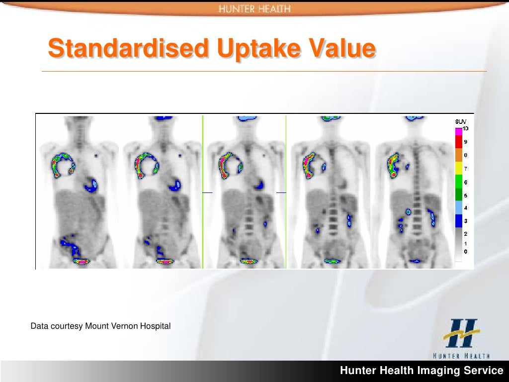
Standardized Uptake Value (SUV) is a crucial metric derived from Positron Emission Tomography (PET) scans. It quantifies the concentration of a radiotracer, typically a glucose analog, within a specific region of the body. This value is essential for assessing the metabolic activity of tissues and plays a pivotal role in characterizing and diagnosing various medical conditions, particularly cancer.
SUV measurements provide a quantitative representation of the radiotracer uptake, offering valuable insights beyond the visual interpretation of PET images. The standardized nature of SUV allows for comparisons across different patients and institutions, facilitating consistent and reliable evaluations of tumor activity. This standardized approach enhances the accuracy and objectivity of PET scans in the diagnosis and management of cancer.
Definition and Significance of SUV
SUV represents the ratio of the metabolic activity within a region of interest (ROI) to the average metabolic activity of the normal tissue. Higher SUV values typically indicate higher metabolic activity, a characteristic often associated with malignant tumors. This quantitative measure complements the anatomical information provided by structural imaging modalities like CT scans, aiding in a more comprehensive assessment of the disease. The significance lies in its ability to provide a quantitative measure of metabolic activity, allowing for comparison across different patients and institutions.
Role of SUV in Characterizing Tumors
SUV values are instrumental in characterizing tumors by reflecting their metabolic activity. A high SUV may suggest an aggressive, rapidly growing tumor, while a low SUV may indicate a less aggressive tumor. Furthermore, variations in SUV values within a tumor can indicate heterogeneity in the tumor’s metabolic activity, which may correlate with different degrees of malignancy or the presence of different cell types. This information is invaluable for guiding treatment decisions, such as surgical resection, chemotherapy regimens, or radiation therapy targeting.
Factors Influencing SUV Values
Several factors influence SUV values, impacting the interpretation of PET scan results. These include the type of radiotracer used, the acquisition parameters of the PET scan, the patient’s hydration status, and the time elapsed since radiotracer administration. Additionally, the presence of inflammatory processes or other metabolically active tissues can influence SUV values, necessitating careful consideration during interpretation. Clinicians must take into account these variables to ensure accurate assessment.
Common Applications of SUV Analysis in Cancer
SUV analysis is widely used in various types of cancer to assess tumor burden, guide treatment planning, and monitor response to therapy. For instance, in lung cancer, SUV can help differentiate between benign and malignant nodules, enabling timely intervention. Similarly, in breast cancer, SUV measurements can assist in staging and identifying areas of high metabolic activity that may require targeted therapies. In melanoma, SUV analysis helps assess the aggressiveness of the tumor and monitor the effectiveness of treatment. SUV analysis can help clinicians make informed decisions about treatment strategies and patient outcomes.
Table of PET Scan Types and SUV Utilization
| PET Scan Type | SUV Application |
|---|---|
| FDG-PET | Widely used for detecting and characterizing various cancers, assessing tumor response to therapy, and staging. SUV values correlate with tumor metabolic activity. |
| 18F-Choline PET | Specifically used for identifying and assessing the metabolic activity of prostate cancer. SUV values reflect the choline uptake by tumor cells. |
| 11C-Methionine PET | Useful for identifying and characterizing tumors, particularly in the brain and other organs. SUV values correlate with the amino acid uptake by tumor cells. |
SUV Measurement Techniques
Standardized Uptake Value (SUV) measurement is crucial in Positron Emission Tomography (PET) scans for quantifying metabolic activity within tissues. Accurate SUV calculations are essential for clinical diagnosis and treatment planning. The precise determination of SUV values allows clinicians to assess the extent and nature of disease processes.
Accurate SUV calculation requires careful attention to various factors, including patient characteristics, imaging parameters, and the specific region of interest (ROI) selected. Understanding the procedures, methods, and equipment involved in SUV measurements is vital for interpreting PET scan results correctly.
Procedures for Calculating SUV Values
The calculation of SUV values involves several steps, beginning with the acquisition of the PET scan data. This data is then processed to generate images that depict the distribution of radioactivity within the body. Subsequently, regions of interest (ROIs) are delineated on these images, encompassing the target tissues or organs of interest. The total radioactivity within the delineated ROI is then measured, and this value is normalized by dividing it by the patient’s weight and the administered dose of radiotracer. This normalization process ensures that SUV values are comparable across different patients and scans.
Methods for Determining SUV
Different methods exist for determining SUV values, each with its own advantages and limitations. The most common approach is to use the maximum SUV (SUVmax), which represents the highest concentration of radiotracer within the ROI. This approach is useful for identifying areas of high metabolic activity. Other methods, such as the mean SUV (SUVmean), can provide a more comprehensive assessment of the overall metabolic activity within the region. The choice of method depends on the specific clinical question and the characteristics of the PET scan data.
Equipment Involved in SUV Measurements
The equipment used in SUV measurements is critical for accurate and reliable results. PET scanners, which are the primary imaging instruments, must be calibrated and maintained to ensure precise measurements. Software applications are also essential for processing the acquired data and calculating SUV values. The software must be accurate and reliable, and its algorithms must be validated to ensure the reliability of the results. Specialized software is crucial for delineating the ROI and performing the calculations. Additionally, patient data, including weight and administered dose, must be accurately recorded and integrated into the calculation process.
Factors Affecting the Accuracy of SUV Measurements
Several factors can influence the accuracy of SUV measurements, including patient factors, scan parameters, and the selection of ROIs. Patient weight, body composition, and hydration status can all affect the concentration of radiotracer within the body, leading to variations in SUV values. Scan parameters, such as the injected dose and the duration of the scan, also play a significant role. Inaccurate delineation of the ROI can lead to inaccurate SUV values. Furthermore, the presence of artifacts in the PET images can also introduce errors in SUV calculations.
SUV Calculation Formulas
| Formula | Description |
|---|---|
| SUVmax = (Maximum radioactivity within ROI) / ((Patient weight in kg) * (injected dose in MBq) / (Body surface area in m2)) | Calculates the maximum standardized uptake value within a region of interest. |
| SUVmean = (Average radioactivity within ROI) / ((Patient weight in kg) * (injected dose in MBq) / (Body surface area in m2)) | Calculates the mean standardized uptake value within a region of interest. |
| SUVpeak = (Radioactivity in the region of peak uptake) / ((Patient weight in kg) * (injected dose in MBq)) | Calculates the peak SUV, focusing on the area of highest metabolic activity. |
The formulas presented above demonstrate the fundamental principles for calculating SUV values. These formulas should be applied according to the specific instructions provided by the PET imaging center and the radiopharmaceutical manufacturer.
Interpretation of SUV Values

Interpreting Standardized Uptake Values (SUV) from Positron Emission Tomography (PET) scans is crucial for evaluating the metabolic activity of tissues, particularly in cancer detection and staging. SUV values, derived from the concentration of radiotracer in a specific region of interest, offer insights into the metabolic characteristics of a lesion, aiding clinicians in determining its malignancy and aggressiveness.
SUV values are not diagnostic in isolation but serve as a valuable tool in conjunction with other imaging modalities and clinical information. A high SUV often suggests increased metabolic activity, potentially indicating malignancy, while a low SUV may point to a benign process. However, a nuanced approach is essential, considering the individual patient’s medical history, symptoms, and other imaging results.
SUV Values and Cancer Detection
SUV values provide a relative measure of metabolic activity. Higher SUV values generally correspond to a greater uptake of the radiotracer, which is often associated with increased cellular activity, such as in rapidly dividing cells found in malignant tumors. Conversely, lower SUV values suggest lower metabolic activity, which might be indicative of a benign process or a less aggressive form of cancer.
Examples of SUV Values and Implications
A patient with a lung nodule exhibiting an SUV of 10 might suggest a higher likelihood of malignancy compared to a nodule with an SUV of 2. However, these values are not absolute indicators; other factors, such as the size and location of the lesion, must be considered. Furthermore, the specific radiotracer used and the scanning protocol can influence SUV values. For instance, a patient with a breast mass showing an SUV of 3 in one scan protocol might have a different SUV value in a different protocol.
Limitations of SUV as a Sole Diagnostic Tool
While SUV values offer valuable insights, relying solely on them for diagnosis is inadequate. Numerous factors can influence SUV values, including patient preparation, injection technique, and scanning parameters. Additionally, SUV values are relative and context-dependent. A high SUV value in a patient with a known history of a benign condition might not necessarily indicate malignancy. Therefore, SUV values should be interpreted in conjunction with other diagnostic modalities, such as biopsy, and clinical findings.
Comparison of High and Low SUV Values
| SUV Value | Tumor Characteristics |
|---|---|
| High SUV (e.g., >10) | Potentially aggressive tumor; increased metabolic activity; may indicate a higher likelihood of malignancy; often seen in rapidly growing tumors; larger tumor size; can be indicative of more active and aggressive cancer cells. |
| Low SUV (e.g., <2) | May indicate a benign lesion; lower metabolic activity; potentially less aggressive; smaller tumor size; slower growth rate. This does not definitively rule out malignancy but suggests a lower likelihood compared to high SUV values. |
SUV and Disease Progression
Changes in Standardized Uptake Values (SUV) on Positron Emission Tomography (PET) scans are crucial indicators of disease progression and response to treatment in oncology. Tracking SUV values over time provides valuable insights into tumor behavior and allows clinicians to assess the effectiveness of various therapies. This allows for more precise and timely adjustments to treatment strategies, ultimately improving patient outcomes.
Understanding how SUV values correlate with disease progression is vital for personalized cancer care. Monitoring SUV changes provides clinicians with a quantitative measure of tumor activity, which is often more reliable than visual assessment alone. This quantitative measure aids in making informed decisions regarding treatment modifications, potentially leading to improved patient outcomes.
SUV Changes Reflecting Disease Progression
SUV values can significantly increase as tumors grow and proliferate. Conversely, a decrease in SUV values often indicates tumor shrinkage or response to treatment. This change in SUV reflects the metabolic activity within the tumor, which is directly related to its growth and spread. The rate of SUV change is often correlated with the rate of tumor growth or regression.
Clinical Significance of Tracking SUV Changes
Tracking SUV changes over time allows clinicians to monitor disease progression and response to treatment with greater precision. This information is particularly valuable in making critical decisions about treatment adjustments. The dynamic nature of SUV values allows for a more nuanced understanding of tumor behavior compared to static imaging data.
SUV in Monitoring Treatment Efficacy
SUV values are a valuable tool in monitoring the efficacy of different cancer therapies. A significant decrease in SUV values following treatment can suggest a positive response, while a stable or increasing SUV value may indicate treatment resistance. This information is critical for adapting treatment plans in real-time. For example, if a patient’s SUV values remain elevated after a period of treatment, it may signal the need for a change in therapy to achieve better results.
Examples of SUV Trends in Patients Undergoing Different Treatments
| Treatment Type | SUV Trend |
|---|---|
| Chemotherapy | A significant decrease in SUV values is often observed in patients responding to chemotherapy. This decrease reflects a reduction in tumor metabolism. Conversely, stable or increasing SUV values may indicate treatment resistance. |
| Targeted Therapy | Patients responding to targeted therapies often show a decrease in SUV values. The extent of the decrease can vary depending on the specific targeted therapy and the patient’s individual response. Stable or increasing SUV values could suggest that the targeted therapy is not effectively inhibiting tumor growth. |
| Radiation Therapy | Radiation therapy can sometimes lead to a decrease in SUV values, indicating a reduction in tumor metabolism. The magnitude of the change varies, depending on the type and extent of radiation therapy administered. Failure to observe a decrease in SUV could indicate that the radiation therapy is not effectively targeting the tumor. |
| Immunotherapy | Immunotherapy’s impact on SUV values can vary significantly between patients. Some patients may show a substantial decrease in SUV values, indicating a positive response. However, others may not show any significant change or may even show an increase in SUV, indicating that the therapy is not effective in controlling tumor growth. |
SUV in Specific Cancer Types
Standardized Uptake Values (SUV) in Positron Emission Tomography (PET) scans provide valuable insights into the metabolic activity of tumors, which can vary significantly across different cancer types. Understanding these variations is crucial for accurate diagnosis, staging, and treatment planning. SUV values, while not a definitive diagnostic tool, are an important component of the overall assessment in conjunction with other imaging and clinical data.
Lung Cancer
Lung cancer, a leading cause of cancer-related deaths globally, exhibits a wide range of SUV characteristics. Small cell lung cancers often show lower SUV values compared to non-small cell lung cancers (NSCLC). However, there can be considerable overlap, making SUV analysis alone insufficient for definitive diagnosis. SUV values can be correlated with tumor aggressiveness, providing an indication of the extent of the disease and the potential for aggressive behavior. For instance, higher SUV values in NSCLC may suggest a more aggressive tumor and a poorer prognosis. Furthermore, SUV changes during treatment can indicate response or resistance.
Breast Cancer
SUV values in breast cancer vary depending on the tumor subtype and stage. For example, some studies suggest that triple-negative breast cancers might exhibit higher SUV values compared to hormone receptor-positive cancers. The SUV values can reflect the metabolic activity of the tumor, which may differ depending on the presence of specific receptors and other biological factors. Furthermore, SUV changes during neoadjuvant chemotherapy can be a useful indicator of treatment response, assisting in guiding treatment decisions.
Colon Cancer
Colon cancer, typically characterized by a slow growth rate, can manifest varying SUV values. The SUV values in colorectal metastases can sometimes be elevated, signifying higher metabolic activity compared to primary tumors. However, SUV values in colon cancer are not always indicative of the severity of the disease. It’s important to note that the SUV values are only one aspect of the overall assessment, which should also include clinical information, pathology reports, and other imaging data.
SUV Patterns and Comparisons
Different cancers exhibit distinct SUV patterns. For instance, in lung cancer, SUV values may vary depending on the histological subtype. In breast cancer, SUV values may reflect the presence of specific receptors, and in colon cancer, SUV values may be higher in metastatic sites compared to the primary tumor. Comparison of SUV patterns across these cancers helps to refine the diagnostic process and provides a clearer understanding of the disease characteristics. The variability in SUV patterns across these cancer types highlights the need for careful consideration of the clinical context and other diagnostic data.
Treatment Decisions Informed by SUV Analysis
SUV values can be valuable tools for guiding treatment decisions in various cancers. For instance, in lung cancer, higher SUV values may suggest a more aggressive tumor requiring more aggressive treatment approaches. In breast cancer, SUV changes during neoadjuvant chemotherapy can be an indicator of response, helping physicians to adjust treatment strategies. This allows for more personalized and effective treatment plans.
Challenges in Interpreting SUV Values
Interpreting SUV values across diverse cancer types presents several challenges. One significant challenge is the variability in SUV values among patients with the same cancer type. Factors like patient age, overall health, and treatment history can influence SUV values, making it difficult to draw definitive conclusions solely based on SUV data. Furthermore, SUV values are not always directly correlated with tumor size or stage. The interpretation of SUV values should always be performed in conjunction with other clinical and imaging data to avoid misdiagnosis or inappropriate treatment decisions.
Typical SUV Ranges for Various Cancer Types
| Cancer Type | Typical SUV Range |
|---|---|
| Lung Cancer (NSCLC) | 2-10 |
| Breast Cancer | 1-8 |
| Colon Cancer | 1-5 |
Note: These are approximate ranges and can vary significantly depending on individual factors and the specific characteristics of the tumor.
SUV and Patient Factors
Standardized Uptake Value (SUV) in Positron Emission Tomography (PET) scans is a crucial tool for assessing tumor activity. However, inherent patient variability can significantly impact SUV measurements. Factors like age, weight, and metabolic rate influence the uptake and distribution of radiotracers, leading to discrepancies in SUV values that need careful consideration during interpretation. Understanding these influences is vital for accurate diagnosis and treatment planning.
Patient Factors Affecting SUV Values
Patient characteristics like age, weight, and metabolic rate directly impact the distribution and metabolism of the radiotracer within the body. Variations in these factors can lead to substantial differences in SUV values, potentially obscuring the true nature of tumor activity. Age-related changes in metabolism, for example, can alter the uptake of the radiotracer, resulting in potentially misrepresented SUV levels. Similarly, differences in body weight can affect the concentration of the radiotracer, influencing the apparent SUV. Understanding these factors allows clinicians to interpret SUV values more accurately, leading to improved diagnostic precision.
Importance of Considering Patient-Specific Factors
Ignoring patient-specific factors during SUV interpretation can lead to misdiagnosis and inappropriate treatment strategies. For example, a high SUV value in an elderly patient might not necessarily indicate aggressive tumor behavior but could simply reflect age-related metabolic differences. Conversely, a low SUV in a patient with a high metabolic rate might be a result of the faster clearance of the radiotracer, not necessarily indicative of a less aggressive tumor. Consequently, the careful consideration of patient-specific variables is crucial for accurate clinical decision-making.
Methods for Correcting SUV Values
Various methods exist for adjusting SUV values to account for patient-specific factors. One common approach involves normalizing SUV values based on patient weight. This involves dividing the SUV by the patient’s weight to obtain a standardized value that accounts for differences in body size. Another method involves incorporating patient age and gender into the normalization equation, leading to more refined adjustments. A sophisticated method could incorporate factors like metabolic rate, activity level, and even pre-existing medical conditions.
Example Adjustments for Different Patient Characteristics
| Patient Factor | Adjustment Method |
|---|---|
| Weight | SUVweight-normalized = SUV / Weight (kg) |
| Age | Adjustments are often incorporated into the normalization equations using age-specific metabolic rate models. The complexity of this adjustment depends on the specific radiotracer and the software used. |
| Metabolic Rate | SUVmetabolic-adjusted = SUV * (Metabolic Rate / Average Metabolic Rate) This method requires estimating or measuring the metabolic rate of the patient. |
| Gender | While gender often plays a role in metabolic rate, it’s less frequently a standalone adjustment. The impact of gender is usually integrated into age-specific metabolic rate models. |
These adjustments help standardize SUV values, enabling more reliable comparisons across different patients, regardless of their individual characteristics.
SUV and Future Directions

Standardized Uptake Value (SUV) in Positron Emission Tomography (PET) scans has revolutionized oncology, enabling non-invasive assessment of tumor characteristics and disease progression. However, the interpretation of SUV values remains a complex process, and future directions aim to enhance the accuracy and reliability of this technique. Advanced methodologies and technological advancements promise to provide a more comprehensive and personalized approach to patient care.
The future of SUV analysis lies in integrating multiple data points from various imaging modalities, including PET, MRI, and CT scans. Combining these diverse datasets can provide a more holistic understanding of the tumor microenvironment and its response to treatment. This integrated approach will allow for more precise characterization of tumor heterogeneity and potential treatment responses, paving the way for personalized treatment strategies.
Advanced Techniques in Improving SUV Analysis
Recent advancements in image processing and machine learning techniques are transforming SUV analysis. Algorithms are being developed to automatically segment tumors, quantify uptake in specific regions, and predict tumor behavior. These automated systems can reduce inter-observer variability and streamline the analysis process, allowing for faster and more consistent interpretations. Machine learning models can also be trained to identify subtle patterns in SUV data that might indicate early signs of treatment resistance or disease recurrence. This can aid clinicians in tailoring treatment plans and potentially improving patient outcomes.
Future Research Directions
Future research in SUV analysis should focus on developing more sophisticated models to account for variations in patient characteristics, such as age, weight, and underlying health conditions. The development of standardized protocols for SUV measurement across different imaging systems is also critical. Furthermore, investigating the relationship between SUV values and specific molecular markers of cancer will help to further refine the interpretation of SUV data. Ultimately, researchers aim to create a more robust and predictive framework for utilizing SUV data in clinical decision-making.
New Technologies and Methods for SUV Analysis
Several novel technologies are emerging to enhance the precision and accuracy of SUV measurements. These include:
- Deep Learning-Based Segmentation: Deep learning algorithms are being trained on large datasets of PET images to automatically segment tumors with greater accuracy than traditional methods. This automation reduces manual effort and improves consistency in SUV quantification.
- Hybrid PET/MRI Imaging: Combining PET and MRI data provides complementary information about tumor morphology and metabolic activity. This integrated approach can offer a more comprehensive picture of the tumor, potentially enhancing the accuracy of SUV analysis and improving diagnostic confidence.
- Multiparametric Analysis: Incorporating other imaging modalities, such as CT, into the analysis can provide a more detailed view of the tumor’s location, size, and surrounding tissues. This can enhance the accuracy of SUV measurements and improve the interpretation of SUV values.
Potential Impact on Patient Care
Advancements in SUV analysis have the potential to significantly impact patient care. More accurate and reliable SUV measurements can aid in earlier detection of cancer, more precise staging, and more effective treatment planning. Personalized treatment strategies based on SUV data could lead to improved outcomes, reduced side effects, and potentially longer survival times.
Summary of Future Trends and Developments
| Trend | Description |
|---|---|
| Automated SUV Analysis | Machine learning algorithms are being developed to automate tumor segmentation and SUV quantification, improving consistency and reducing inter-observer variability. |
| Hybrid Imaging Modalities | Combining PET with other imaging modalities, such as MRI and CT, provides a more comprehensive understanding of the tumor microenvironment, leading to more accurate SUV interpretation. |
| Multiparametric Analysis | Integration of SUV data with other clinical and molecular data, such as genetic profiles, will further refine the understanding of SUV and its relationship to disease progression. |
| Personalized Treatment Strategies | SUV data, combined with other factors, can help tailor treatment plans for individual patients, potentially improving outcomes and reducing adverse effects. |