Table of contents:
Introduction to SUV 5.2 PET Scan
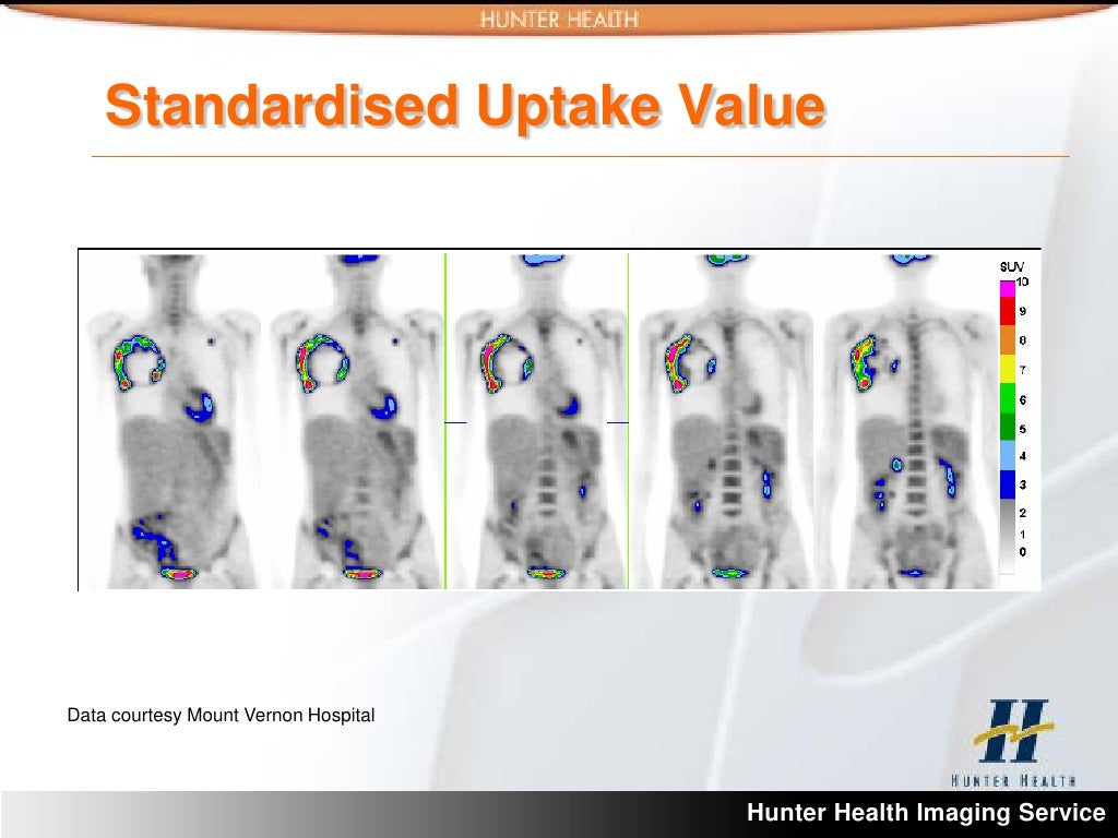
Positron emission tomography (PET) scans are powerful diagnostic tools used in medicine to visualize metabolic activity within the body. A 5.2 SUV (standardized uptake value) PET scan is a specific type of PET scan that measures the concentration of a radiotracer, usually a glucose analog, in tissues. This concentration is then standardized to a baseline value, providing a quantitative measure of metabolic activity. Understanding the 5.2 SUV value is crucial for diagnosing various conditions, particularly in oncology and other areas of medicine.
The purpose of a 5.2 SUV PET scan is to identify areas of high metabolic activity, which can indicate the presence of cancer or other abnormalities. The applications extend beyond oncology, encompassing various fields like neurology, cardiology, and infectious disease diagnosis. This advanced imaging technique offers a non-invasive method for evaluating organ function and identifying potential problems at an early stage.
Technical Principles of PET Scans
PET scans utilize radioactive tracers that emit positrons, which then annihilate with electrons in the body. This annihilation process produces two gamma rays traveling in opposite directions. These gamma rays are detected by specialized detectors surrounding the patient. The location and intensity of the detected gamma rays are used to create images that represent the distribution of the radiotracer within the body. The 5.2 SUV value is derived from the concentration of the radiotracer in a specific region of interest, compared to a standard uptake value (typically the average uptake in the liver or blood pool).
Patient Preparation for a 5.2 SUV PET Scan
Patients undergoing a 5.2 SUV PET scan typically require fasting for a period before the procedure, often ranging from 4 to 6 hours. This fasting period is to ensure that the glucose levels in the body are relatively stable. Patients should also inform their physicians about any medications they are currently taking, as some medications can interfere with the results. Specific dietary instructions and medication adjustments may vary depending on the patient’s individual condition and the specific protocol followed. This preparation ensures the accuracy of the scan by minimizing the influence of external factors.
Types of SUV PET Scans
Different types of PET scans are tailored to address specific needs. Whole-body scans provide a comprehensive overview of metabolic activity throughout the body. Targeted region scans focus on specific organs or areas of interest, allowing for a more detailed analysis of metabolic activity in those regions. The choice of scan type depends on the clinical question being investigated. Examples include scans of the brain to detect neurological conditions or scans of the abdomen to evaluate potential tumors.
Factors Influencing SUV Values
Standardized Uptake Value (SUV) in Positron Emission Tomography (PET) scans provides a crucial metric for evaluating metabolic activity in tissues. Understanding the factors that influence SUV values is essential for accurate interpretation and clinical decision-making. Variability in SUV measurements can arise from several sources, including patient-specific characteristics and the scanning process itself.
Accurate interpretation of SUV values is critical for detecting and characterizing abnormalities. Factors like hydration, recent meals, and the injected radiotracer can significantly impact SUV measurements. This detailed analysis will highlight these influences, offering insights into how these variables affect SUV values, and consequently, the diagnostic interpretation of PET scans.
Impact of Physiological Conditions
Physiological conditions, such as hydration and recent meal intake, significantly affect SUV values. Dehydration can lead to higher SUV values as the concentration of the radiotracer increases within the tissues. Conversely, excessive hydration can dilute the tracer, potentially lowering SUV values. Similarly, recent meals, particularly those high in carbohydrates, can increase glucose uptake and thus lead to elevated SUV values in glucose-avid tissues. These effects can confound interpretation, and clinicians must consider these physiological factors when assessing SUV values.
Influence of Radiotracer Characteristics
The radiotracer used in the PET scan plays a pivotal role in SUV measurements. Different radiotracers have varying characteristics, including their half-life, metabolism, and tissue uptake. The radiotracer’s specific properties directly impact how it’s distributed in the body, influencing SUV values. For example, a radiotracer with a longer half-life might result in a more prolonged elevation of SUV values in the target tissue compared to one with a shorter half-life.
Variations Across Patient Populations
SUV values can differ across various patient populations, including those categorized by age and gender. Age-related changes in metabolism and tissue composition can affect radiotracer uptake, resulting in different SUV values compared to younger individuals. Similarly, gender differences in metabolic rates might influence SUV values. It’s crucial to consider these potential variations when comparing SUV values across different patient cohorts.
Examples of Abnormal SUV Values and Implications
Abnormal SUV values, either elevated or decreased, can provide valuable insights into potential pathologies. Elevated SUV values in a particular area may indicate increased metabolic activity, potentially suggesting malignancy or inflammation. For example, a high SUV value in a lung region might suggest lung cancer. Conversely, decreased SUV values may signify reduced metabolic activity, potentially pointing towards tissue atrophy or ischemia. For instance, a low SUV value in a brain region might indicate a stroke or other neurological conditions. Careful consideration of the clinical context and other imaging findings is essential for accurate interpretation.
Interpretation and Analysis of SUV Results

Analyzing SUV (standardized uptake value) results from PET scans requires a meticulous approach, combining quantitative data with clinical context. Radiologists and other medical professionals use standardized methods to interpret SUV values, ensuring accurate assessment of the presence, extent, and characteristics of metabolic activity within the body. This process involves comparing individual SUV values to established reference ranges and considering factors like patient demographics, scan protocol, and potential confounding variables.
Interpreting SUV values is not simply a matter of comparing a number to a table; it demands a comprehensive understanding of the patient’s medical history, the purpose of the scan, and the potential implications of the findings. Radiologists carefully evaluate the spatial distribution and intensity of SUV uptake, noting any unusual patterns or focal areas of increased activity. This careful consideration ensures that the information gleaned from the scan is correctly contextualized and used to inform diagnostic and therapeutic decisions.
Typical Methods for Analyzing SUV PET Scan Results
Radiologists employ a variety of methods to analyze SUV PET scan results. These methods include visual assessment, semi-quantitative analysis, and fully quantitative analysis. Visual assessment involves a subjective evaluation of the scan images, identifying areas of increased or decreased uptake. Semi-quantitative analysis involves measuring SUV values in specific regions of interest (ROIs) and comparing them to reference ranges. Fully quantitative analysis, often using sophisticated software, considers a wider range of factors and generates more detailed reports.
Comparison to Normal Ranges
A crucial aspect of SUV analysis is comparing the obtained values to established normal ranges. These ranges are usually specific to the organ or tissue being evaluated and are influenced by various factors such as age, sex, and underlying health conditions. These ranges are not absolute; they represent a statistical average, and individual variations are expected. Deviations from these ranges, whether elevated or depressed, can suggest abnormalities, warranting further investigation.
Typical SUV Values for Different Organs and Tissues
| Organ/Tissue | Typical SUV Values (mean ± SD) |
|---|---|
| Brain | 0.5-2.5 |
| Heart | 0.8-1.8 |
| Liver | 2.0-5.0 |
| Kidney | 1.0-3.5 |
| Skeletal Muscle | 0.5-1.5 |
| Lung | 0.5-2.0 |
Note: These values are approximate and can vary based on the specific PET scan protocol and patient characteristics. Consult the specific guidelines and reports generated by the imaging center for detailed reference ranges.
Limitations of Using SUV Values for Diagnosis
While SUV values provide valuable information, they have limitations. They are not diagnostic in themselves and should be considered alongside other clinical data. Factors such as patient preparation, scan protocol, and individual physiological variations can influence SUV values, potentially leading to inaccurate interpretations. For instance, inflammation or hydration levels can impact uptake, leading to false positives or negatives. SUV values should be interpreted in conjunction with other imaging modalities, clinical history, and laboratory results. The overall clinical context is essential for accurate and meaningful interpretation.
Clinical Applications and Case Studies

5.2 SUV PET scans have emerged as a powerful tool in the diagnosis and management of various medical conditions, particularly in oncology. This advanced imaging technique provides crucial information about metabolic activity within tissues, enabling clinicians to differentiate between benign and malignant processes, assess the extent of disease, and monitor treatment response. This section will delve into the clinical applications of 5.2 SUV PET scans in diverse medical contexts, highlighting their role in cancer detection, staging, and treatment monitoring.
The ability of 5.2 SUV PET scans to identify areas of increased metabolic activity within the body allows for early detection of potentially cancerous lesions. Furthermore, the quantitative nature of SUV values facilitates precise staging of tumors, aiding in determining the extent of disease spread and its impact on surrounding organs. This detailed information is critical for developing personalized treatment plans and predicting patient outcomes.
Clinical Applications in Oncology
5.2 SUV PET scans are invaluable in the detection and staging of various cancers. The scans provide a non-invasive method to assess the metabolic activity of tumors, which often differs significantly from that of surrounding healthy tissue. This allows for a more precise assessment of the extent of disease and its potential impact on surrounding organs. High SUV values typically correlate with more aggressive cancers.
Cancer Detection and Staging
The high sensitivity and specificity of 5.2 SUV PET scans contribute significantly to cancer detection and staging. In comparison to traditional imaging techniques like CT scans, 5.2 SUV PET scans provide a more comprehensive evaluation of tumor metabolic activity. This improved assessment aids in identifying early-stage cancers, potentially enabling more effective and less invasive treatments. For example, in cases of suspected lung cancer, a 5.2 SUV PET scan can reveal areas of abnormal metabolic activity that might not be visible on a CT scan, thereby facilitating earlier diagnosis.
Case Study: Diagnosing Metastatic Disease
A 65-year-old male patient presented with persistent back pain and fatigue. Initial blood tests and imaging (CT scan) were inconclusive. A 5.2 SUV PET scan revealed a focal area of increased metabolic activity in the lumbar spine, with SUV values significantly higher than the surrounding tissue. Further investigation confirmed the presence of a malignant tumor, with evidence of metastatic spread to the spine. This case exemplifies the utility of 5.2 SUV PET scans in detecting and characterizing metastatic disease, enabling timely intervention and potentially improving patient outcomes.
Differentiating Benign and Malignant Lesions
Differentiation between benign and malignant lesions is a crucial aspect of cancer diagnosis. 5.2 SUV PET scans, by assessing metabolic activity, can often provide valuable insights. Benign lesions typically exhibit lower SUV values compared to malignant lesions, which tend to demonstrate higher values. This difference in metabolic activity aids clinicians in differentiating between benign and malignant conditions, guiding further diagnostic procedures and treatment strategies. For instance, a suspicious nodule in the breast with a high SUV value on a 5.2 SUV PET scan would suggest malignancy, prompting further investigation.
Clinical Scenarios Utilizing 5.2 SUV PET Scans
| Clinical Scenario | Benefits of 5.2 SUV PET Scan |
|---|---|
| Suspected recurrence of lymphoma | Detects residual disease and assesses its metabolic activity, aiding in treatment planning. |
| Monitoring response to cancer therapy | Evaluates the effectiveness of treatment by assessing changes in SUV values over time. |
| Staging of primary lung cancer | Accurately determines the extent of the disease, including lymph node involvement. |
| Evaluating suspected metastasis | Identifies areas of increased metabolic activity that might indicate the spread of cancer. |
| Differentiating between benign and malignant tumors | Provides a quantitative measure of metabolic activity, aiding in the diagnosis of suspicious lesions. |
Image Analysis and Reporting
Image analysis plays a critical role in the interpretation of 5.2 SUV PET scans, providing crucial information for diagnosis and treatment planning. Accurate and comprehensive image analysis requires a standardized approach to ensure consistency and reliability across different institutions. This section details the process of acquiring and analyzing 5.2 SUV PET scan images, including the various formats used and the methods employed to interpret SUV values.
The standardized reporting of 5.2 SUV PET scan images ensures that clinicians across various settings can interpret the results consistently and effectively. This consistency is vital for accurate diagnosis and treatment planning. Different image analysis methods and software tools are utilized to extract relevant information from the images, providing a comprehensive view of the metabolic activity within the body.
Image Acquisition and Presentation
The process of obtaining images from a 5.2 SUV PET scan typically involves the acquisition of a series of two-dimensional images, often encompassing multiple anatomical planes. These images are typically captured using specialized PET scanners, and the quality and resolution of the images are crucial for accurate analysis. The patient’s position during the scan is carefully monitored to minimize motion artifacts, which can compromise the quality of the data. Data acquisition parameters are meticulously controlled to ensure the reproducibility and reliability of the scan results.
Image Formats for Reporting
PET scan images are typically presented in three primary formats: axial, coronal, and sagittal. Axial images display the anatomical structures in a cross-sectional view, providing a detailed view of the target organ or area of interest. Coronal images present the structures in a frontal view, while sagittal images offer a side-view perspective. The choice of which view to utilize is based on the specific anatomical area or pathology being evaluated.
- Axial Images: These are the most common view for initial evaluation, offering a comprehensive overview of the target area. Clinicians often use axial images to identify the presence and extent of abnormal metabolic activity. This view allows for a detailed assessment of structures in the transverse plane, providing insight into the spatial distribution of the metabolic processes.
- Coronal Images: These images provide a frontal perspective, offering valuable insights into the anatomical structures in a sagittal plane. They are particularly useful for evaluating lesions or abnormalities that extend along the frontal axis. The anatomical orientation in this view allows for a thorough assessment of lesions that extend across the coronal plane.
- Sagittal Images: Sagittal images provide a side-view perspective of the structures, which are essential for evaluating lesions or abnormalities that extend along the sagittal axis. These images are particularly valuable in assessing the anatomical relationships of lesions in the axial plane.
Image Analysis Methods
A variety of methods are used to interpret SUV values from the images. These methods include visual assessment, semi-quantitative analysis, and quantitative analysis, each contributing to a comprehensive evaluation. Visual assessment involves the careful examination of the images to identify areas of increased or decreased metabolic activity. Semi-quantitative analysis employs a scoring system or visual grading scale to estimate the intensity of metabolic activity, while quantitative analysis uses sophisticated software and algorithms to calculate and measure SUV values in a precise manner.
Image Analysis Techniques for Different Scan Types
| Scan Type | Image Analysis Techniques |
|---|---|
| Whole-body PET | Visual assessment of areas of increased uptake, followed by region-of-interest (ROI) analysis to quantify SUV values. This is often coupled with anatomical correlation from co-registered CT or MRI images. |
| Targeted PET | Detailed ROI analysis to precisely quantify SUV values in specific anatomical regions of interest. This approach often relies on anatomical templates or co-registration with anatomical images. |
| Dynamic PET | Time-activity curves (TACs) are generated and analyzed to assess the kinetic properties of the radiotracer. This can provide insight into the biological mechanisms underlying the observed metabolic activity. Analysis may also include calculation of the uptake rate constant or the washout rate constant to evaluate metabolic kinetics. |
Comparison with Other Imaging Techniques
5.2 SUV PET scans provide valuable insights into metabolic activity, but their interpretation is often enhanced by comparison with other imaging modalities. Understanding the strengths and weaknesses of each technique, as well as the synergistic benefits of combined imaging, is crucial for accurate diagnosis and treatment planning. This section delves into the comparative analysis of 5.2 SUV PET scans with computed tomography (CT), magnetic resonance imaging (MRI), and other relevant modalities.
CT and MRI are established imaging techniques with distinct advantages. CT excels at anatomical detail, providing high-resolution images of bone and soft tissues, while MRI offers superior soft tissue contrast, particularly for visualizing the intricacies of the nervous system and musculoskeletal structures. However, both modalities lack the metabolic information provided by PET scans.
Comparison of SUV PET with CT
CT scans are widely used for anatomical delineation, particularly in cases of suspected malignancy, trauma, or vascular disease. Their high spatial resolution allows for precise localization of lesions. 5.2 SUV PET scans, on the other hand, provide metabolic information, allowing assessment of tumor activity and metabolic changes. The combination of high-resolution anatomical details from CT and the metabolic insights from PET can significantly improve diagnostic accuracy.
Comparison of SUV PET with MRI
MRI is a powerful tool for visualizing soft tissues, enabling detailed assessment of brain, spinal cord, and musculoskeletal structures. It excels in differentiating various soft tissue types and identifying subtle pathologies. While MRI is superior for soft tissue imaging, it lacks the ability to directly assess metabolic activity like 5.2 SUV PET scans. The complementary nature of these modalities allows for a more comprehensive understanding of the disease process.
Combined Imaging Approaches
The integration of multiple imaging techniques, often referred to as multi-modality imaging, can provide a more comprehensive understanding of a disease than any single modality alone. Combining 5.2 SUV PET scans with CT or MRI allows clinicians to fuse anatomical information with metabolic data, enabling better localization and characterization of disease. This integrated approach is particularly valuable in oncology, where the combined data aids in determining the extent of disease, assessing treatment response, and monitoring disease progression.
Situations Favoring SUV PET Scans
5.2 SUV PET scans are particularly beneficial in scenarios where metabolic activity plays a crucial role in disease diagnosis and prognosis. For example, in oncology, identifying areas of high metabolic activity within a tumor can aid in staging and treatment planning. In neurology, 5.2 SUV PET scans can highlight metabolic changes associated with certain neurological disorders.
Value of Multi-Modality Imaging
| Imaging Modality | Strengths | Weaknesses | Synergistic Value with 5.2 SUV PET |
|---|---|---|---|
| CT | Excellent anatomical detail, high spatial resolution, fast acquisition | Limited soft tissue contrast | Provides anatomical context for interpreting metabolic findings from PET |
| MRI | Superior soft tissue contrast, detailed visualization of soft tissues and organs | Longer acquisition time, potential for artifacts | Provides detailed soft tissue information complementary to PET metabolic assessment |
| 5.2 SUV PET | Provides metabolic information, assesses tumor activity, and highlights areas of high metabolic activity | Limited anatomical detail, may need co-registration with other modalities for precise localization | Provides essential metabolic information for characterizing lesions and guiding treatment |
Future Directions and Emerging Technologies
The field of 5.2 SUV PET scans is rapidly evolving, driven by advancements in technology and a growing need for more accurate and informative diagnostic tools. This section explores potential future developments, focusing on the integration of artificial intelligence, novel radiotracers, and emerging research areas to enhance the utility and precision of this valuable imaging modality.
Potential Advancements in 5.2 SUV PET Scan Technology
Ongoing research aims to refine the acquisition and processing of PET scan data. This includes improvements in detector technology for enhanced sensitivity and resolution, allowing for clearer visualization of metabolic activity and reducing radiation exposure. Furthermore, advancements in image reconstruction algorithms promise to improve image quality, reducing artifacts and enhancing the delineation of metabolically active regions. These refinements will lead to more precise quantitative measurements of SUV values and improved diagnostic accuracy.
Role of Artificial Intelligence in Improving Image Analysis and Interpretation
Artificial intelligence (AI) is poised to revolutionize the interpretation of 5.2 SUV PET scan data. AI algorithms can analyze vast datasets of PET scan images, identifying patterns and anomalies that may be missed by human observers. This capability can accelerate the diagnostic process and potentially lead to earlier and more accurate diagnoses, particularly in complex cases. Machine learning models can be trained to distinguish between benign and malignant lesions based on their metabolic profiles, enhancing the diagnostic confidence and reducing the need for invasive procedures.
Examples of New Radiotracers and Their Potential Applications
The development of novel radiotracers is critical for expanding the applications of 5.2 SUV PET scans. Radiotracers targeting specific molecular targets within tumors or tissues can provide detailed information about disease mechanisms and potentially allow for personalized treatment strategies. For instance, radiotracers designed to bind to specific proteins overexpressed in cancer cells can enhance the ability to distinguish between different types of cancer or stages of disease. This personalized approach allows for tailored treatment plans based on the specific metabolic characteristics of the disease.
Research Areas Related to 5.2 SUV PET Scans
Several research areas are actively exploring the potential of 5.2 SUV PET scans. Studies are focusing on the development of novel radiotracers for various diseases, including neurodegenerative disorders, cardiovascular diseases, and infectious diseases. The use of PET scans to assess treatment response in various cancers is another critical area of research. Furthermore, research into standardizing SUV measurements across different scanners and institutions is vital for ensuring the reproducibility and comparability of results.
Emerging Trends in 5.2 SUV PET Scans
| Trend | Description | Impact |
|---|---|---|
| AI-driven image analysis | Machine learning algorithms are being integrated to automate image analysis, leading to faster and more accurate diagnoses. | Improved diagnostic efficiency, reduced human error, and potential for earlier detection. |
| Development of targeted radiotracers | New radiotracers are being developed to target specific molecular markers in diseases. | Enhanced ability to differentiate between diseases, improved understanding of disease mechanisms, and potential for personalized medicine. |
| Standardization of SUV measurement | Efforts are underway to create standardized protocols for SUV measurements across different institutions. | Improved comparability of results across studies and institutions, increasing the reliability of diagnostic interpretations. |
| Integration with other imaging modalities | Combining PET scans with other imaging techniques like MRI or CT scans provides a more comprehensive view of the disease. | More complete characterization of the disease, aiding in treatment planning and monitoring. |