Introduction to SUV 5.8 PET Scan

A 5.8-liter SUV engine, typically found in large sport utility vehicles, is a high-performance engine, often incorporating sophisticated engineering features to optimize power and efficiency. These vehicles are frequently used for off-roading and towing, requiring substantial power output. The sheer size and weight of these vehicles often necessitate the use of robust mechanical components and specialized designs. This makes them a fascinating subject for comparison with PET scan technologies.
Positron Emission Tomography (PET) scans are a sophisticated medical imaging technique that uses radioactive tracers to visualize metabolic activity within the body. PET scans provide unique insights into organ function and highlight areas of abnormal metabolic activity, which can be critical in diagnosing a variety of medical conditions. The use of radioactive tracers allows medical professionals to visualize the dynamic processes occurring within the body, going beyond the static images provided by other imaging techniques.
While seemingly disparate fields, there is potential overlap between the study of high-performance SUV engines and the analysis of metabolic activity in the body through PET scans. For example, the precise measurement of fuel efficiency in SUVs and the quantification of glucose uptake in PET scans both rely on the careful analysis of measurable data. The meticulous mapping of component performance in an engine can be compared to the detailed mapping of metabolic processes in the body. Furthermore, both require sophisticated instrumentation and rigorous data analysis.
Potential Medical Applications
A range of medical applications can leverage the insights from both SUV performance analysis and PET scans. For example, monitoring the effectiveness of treatments for metabolic disorders can be facilitated by comparing the patient’s PET scan results to the expected metabolic rate. Similarly, the study of cardiovascular disease can benefit from the comparison of PET scan findings on glucose uptake in the heart to engine performance metrics related to fuel efficiency.
Comparison of PET Scan Imaging Techniques
| Imaging Technique | Description | Strengths | Limitations |
|---|---|---|---|
| Fluorodeoxyglucose (FDG) PET | Uses FDG, a radioactive glucose analog, to visualize glucose metabolism. | Widely available, good sensitivity for detecting cancer and other metabolic disorders. | May not be suitable for all tissues or conditions; requires careful interpretation of results. |
| Fluorine-18 (18F) PET | Employs 18F-labeled compounds for imaging specific molecular targets. | High specificity for certain molecules, allowing for targeted imaging. | More specialized, potentially less accessible than FDG PET. |
| Carbon-11 (11C) PET | Utilizes 11C-labeled compounds for imaging specific molecular targets, with a shorter half-life. | Provides rapid imaging of metabolic processes, useful in dynamic studies. | Shorter half-life means more specialized facilities and rapid patient workflow required. |
The table above Artikels some common PET scan imaging techniques. Each technique has its own set of advantages and disadvantages, making careful selection crucial for specific applications. Choosing the correct technique for a given situation ensures the most accurate and relevant results.
SUV 5.8 and PET Scan
Positron emission tomography (PET) scans are powerful diagnostic tools used to visualize metabolic activity within the body. Combining a PET scan with the standardized uptake value (SUV) calculation provides valuable insights into tissue function, aiding in various medical diagnoses. This section delves into the technical aspects of PET scans, focusing on the mechanics, procedures, and the relationship between SUV 5.8 and metabolic activity.
The SUV value, specifically SUV 5.8, is derived from a PET scan and serves as a quantitative measure of tissue metabolic activity. A higher SUV value often correlates with increased metabolic activity, which can be indicative of cancerous or inflammatory processes. This relationship is crucial in understanding the information gleaned from a PET scan and interpreting the SUV 5.8 value.
Mechanics of a PET Scan
PET scans utilize radiotracers, which are short-lived radioactive isotopes that emit positrons. When a positron collides with an electron, two gamma rays are emitted in opposite directions. These gamma rays are detected by specialized detectors surrounding the patient. The precise location and intensity of the emitted gamma rays are used to create a 3D image of the metabolic activity within the body.
Process of a PET Scan
The process of a PET scan involves several key steps. Patients are typically given an intravenous injection of a radiotracer, which allows the radioactive substance to circulate throughout the body. After a specific waiting period, typically determined by the radiotracer used, the PET scan is performed. During the scan, the patient lies on a table that moves through the scanner, allowing the detectors to capture the gamma rays. The scanner collects data that is then processed to create a detailed image.
Relationship Between SUV Uptake and Metabolic Activity
The SUV value is a ratio calculated from the measured signal intensity of a region of interest (ROI) and the injected dose of radiotracer. A higher SUV uptake generally indicates a higher level of metabolic activity in that tissue. This increased activity can be associated with a range of conditions, including inflammation, infection, and tumor growth. For example, a tumor often exhibits increased metabolic activity compared to healthy tissue, resulting in a higher SUV value in the tumor area.
Types of Radiotracers Used in PET Scans
Various radiotracers are used in PET scans, each designed to target specific metabolic pathways or molecules. Common radiotracers include fluorodeoxyglucose (FDG), which is commonly used for cancer detection due to its affinity for glucose metabolism. Other radiotracers can target different metabolic pathways and are used for specific purposes.
Factors Influencing SUV 5.8 Value
The SUV 5.8 value is not a static measure and can be affected by several factors. These factors include the injected dose of radiotracer, the type of radiotracer used, the scanning parameters, and the patient’s physiological state. A table outlining these factors is presented below.
| Factor | Description | Impact on SUV 5.8 |
|---|---|---|
| Injection Dose | Amount of radiotracer administered | Higher dose generally results in a higher SUV value. |
| Radiotracer Type | Specific radiotracer used | Different radiotracers have varying uptake characteristics. |
| Scanning Parameters | Scanner settings and acquisition protocols | Changes in these settings can affect the measured signal. |
| Patient’s Physiological State | Factors like hydration, recent meals, and medications | These factors can influence the uptake and distribution of the radiotracer. |
| Region of Interest (ROI) Definition | Area of the body being analyzed | Precise ROI definition affects the SUV calculation. |
Potential Applications and Uses
The SUV 5.8 PET scan, a specialized imaging technique, offers valuable insights into metabolic activity within the body. Understanding its applications and potential uses is crucial for its effective integration into clinical practice. This section will delve into the diverse range of medical scenarios where an SUV 5.8 PET scan can be a powerful diagnostic tool.
The ability to quantify metabolic activity with high precision makes the SUV 5.8 PET scan a valuable diagnostic tool in various medical fields. This detailed analysis allows for the identification of subtle metabolic changes that might be missed by other imaging techniques, potentially leading to earlier and more accurate diagnoses.
Medical Applications of SUV 5.8 PET Scans
The SUV 5.8 PET scan is a valuable diagnostic tool for various medical conditions. Its ability to quantify metabolic activity allows for the detection of abnormalities in a wide range of tissues and organs. This makes it particularly useful for identifying cancerous lesions and assessing the extent of disease spread.
Diagnosing Various Diseases with PET Scans
PET scans, a crucial imaging technique, are used in diagnosing various diseases. By visualizing metabolic activity, PET scans can detect areas of increased or decreased activity, which can be indicative of various diseases, including cancer, neurodegenerative disorders, and inflammatory conditions. The sensitivity and specificity of PET scans make them valuable in differentiating between benign and malignant conditions.
Accuracy Comparison with Other Imaging Techniques
While PET scans are powerful diagnostic tools, their accuracy needs to be considered in comparison to other imaging techniques. Factors like the specific disease being evaluated and the quality of the scan itself can influence the accuracy of the results. For instance, in detecting certain types of cancer, PET scans might offer higher accuracy than CT scans, while for evaluating bone fractures, X-rays might be more appropriate.
Advantages and Disadvantages of SUV 5.8 PET Scans
The SUV 5.8 PET scan offers several advantages, including its ability to visualize metabolic activity and its high sensitivity for detecting certain types of cancers. However, it also has some disadvantages, such as its relatively high cost and potential for false positives. Careful interpretation of results and consideration of other clinical factors are crucial.
Specific Medical Scenarios
The SUV 5.8 PET scan can be particularly useful in specific medical scenarios. For example, in patients suspected of having metastatic cancer, the scan can help identify the presence and extent of the spread of the disease. It can also be used in monitoring the response to treatment, allowing physicians to assess the effectiveness of therapy in reducing metabolic activity in tumors. Further, in cases of suspected neurodegenerative diseases, PET scans can provide valuable information about the extent of neuronal damage. For example, in patients with Alzheimer’s disease, PET scans can detect amyloid plaques in the brain, supporting the diagnosis.
Interpreting Results and Data Analysis
Interpreting the results of an SUV 5.8 PET scan requires careful consideration of various factors and limitations. The standardized uptake value (SUV) is a crucial metric, reflecting the tissue’s metabolic activity. Accurate interpretation aids in diagnosing and staging diseases, guiding treatment decisions, and monitoring response to therapy. However, a nuanced understanding of the potential confounding factors is essential to avoid misinterpretations.
Factors Affecting SUV 5.8 PET Scan Results
Several factors can influence the SUV values obtained from a PET scan. Patient preparation, including fasting requirements and hydration status, can affect the results. The time elapsed between the injection of the radiotracer and the scan acquisition also plays a significant role. Different scanners and imaging protocols can produce varying SUV values. Furthermore, the presence of other medical conditions, such as diabetes or inflammatory processes, can impact SUV readings. Variations in patient size and body composition also contribute to potential variations in SUV values.
Limitations of SUV 5.8 PET Scan Results
SUV 5.8 PET scans provide valuable information, but they are not without limitations. The SUV value itself does not definitively diagnose a condition. It’s merely an indicator of metabolic activity. Furthermore, the results are relative and depend on the baseline activity of the surrounding tissues. The accuracy of SUV values can be affected by the heterogeneity of the metabolic activity within the lesion or the presence of artifacts in the scan. Lastly, differences in scanner calibration and image processing algorithms can lead to variability in the reported SUV values.
Normal Range of SUV Values
The normal range for SUV values varies significantly depending on the specific region of the body being examined. For example, the brain typically exhibits lower SUV values compared to the liver or kidneys. The normal range is also influenced by the patient’s age, sex, and overall health. Specific ranges should be determined by consulting appropriate medical databases and guidelines, considering the specific anatomical region of interest.
Stages of SUV 5.8 PET Scan Analysis
The analysis of an SUV 5.8 PET scan involves several key stages.
| Stage | Description |
|---|---|
| Data Acquisition and Processing | This involves the collection of PET scan data and its subsequent processing to create images. Critical steps include quality control of the acquired images and correction for artifacts. |
| SUV Calculation | The standardized uptake value (SUV) is calculated for each region of interest (ROI) in the image. This calculation takes into account the injected dose of the radiotracer, the patient’s weight, and the measured radioactivity in the ROI. |
| Region of Interest (ROI) Delineation | This crucial step involves carefully defining the regions of interest within the scan where metabolic activity is to be evaluated. Accurate ROI delineation is critical for accurate SUV calculation. |
| Interpretation and Reporting | The calculated SUV values are interpreted in the context of the patient’s clinical presentation, medical history, and other diagnostic information. The findings are documented in a report that highlights areas of abnormal activity. |
Ethical Considerations and Safety Protocols
SUV 5.8 PET scans, while offering valuable diagnostic insights, raise important ethical and safety concerns that must be carefully addressed. The potential for misinterpretation of results, leading to unnecessary or inappropriate interventions, necessitates a thorough understanding of the limitations of the technology. Equally crucial is ensuring patient safety and well-being throughout the entire scanning process.
Understanding the ethical implications and stringent safety protocols surrounding SUV 5.8 PET scans is paramount. This section delves into the considerations that must be taken into account to ensure responsible and ethical application of this technology, protecting both the patient and the healthcare provider.
Ethical Implications of SUV 5.8 PET Scans
The interpretation of SUV 5.8 values necessitates careful consideration of potential biases and the need for comprehensive clinical context. Uncritical reliance on SUV 5.8 values alone can lead to misdiagnosis and inappropriate treatment plans. Clinicians must exercise sound judgment, considering the patient’s overall health history, symptoms, and other diagnostic findings alongside SUV 5.8 data. Transparency in communicating the limitations of the technology and the importance of a multi-faceted approach to diagnosis is crucial. Patients should be informed about the potential benefits and risks, enabling them to make informed decisions regarding their care.
Safety Protocols for Performing an SUV 5.8 PET Scan
Adherence to strict safety protocols is essential to minimize potential risks associated with PET scans. These protocols encompass radiation safety measures, patient preparation, and post-scan monitoring. Radiation exposure from PET scans should be minimized through optimized scan parameters and appropriate shielding techniques.
- Radiation Dose Minimization: Optimized scan protocols, including reduced scan duration and careful selection of acquisition parameters, are critical to minimize radiation exposure to the patient. Experienced technicians and radiologists must carefully balance image quality with radiation dose to achieve the best possible diagnostic results with the lowest potential risk.
- Patient Preparation: Proper patient preparation is paramount to ensure accurate results and minimize potential complications. This includes fasting guidelines, medication adjustments, and any necessary medical evaluations to ensure the patient is in the best possible physical condition before undergoing the scan.
- Allergy Management: Allergies to contrast agents should be carefully assessed before the scan. Proper measures must be in place to manage any allergic reactions that might occur. Pre-scan medical history reviews are vital to identify and address any potential risks.
- Monitoring Procedures: Post-scan monitoring is essential to detect and address any immediate adverse effects. This includes vital sign checks, observation for any allergic reactions, and appropriate management of potential side effects.
Potential Risks and Side Effects
While generally safe, PET scans, like any medical procedure, carry potential risks and side effects. These risks are typically low, but awareness and appropriate precautions are crucial. Common side effects include mild nausea, fatigue, or discomfort at the injection site. In rare cases, more severe allergic reactions or radiation-related complications can occur.
- Radiation Exposure: PET scans involve exposure to ionizing radiation. While the doses are generally low, cumulative exposure should be considered over time. This is particularly important for patients requiring frequent or multiple scans.
- Allergic Reactions: Some patients may experience allergic reactions to the radiotracer. This is typically managed through pre-scan assessments and appropriate medication protocols.
- Infection: As with any invasive procedure, there’s a theoretical risk of infection at the injection site, but these risks are minimized with proper aseptic technique.
Importance of Patient Preparation
Thorough patient preparation is crucial for a successful and safe SUV 5.8 PET scan. Accurate and reliable results depend on the patient following pre-scan instructions precisely. Proper patient preparation reduces the likelihood of complications and ensures optimal image quality. Patients should be well-informed about the procedures and any necessary precautions to take before, during, and after the scan.
| Safety Precautions | Potential Complications |
|---|---|
| Radiation dose optimization | Radiation-induced effects (rare) |
| Strict adherence to fasting guidelines | Nausea, vomiting (rare) |
| Careful allergy assessment | Allergic reactions (rare) |
| Post-scan monitoring | Discomfort at injection site, fatigue (common) |
| Proper injection technique | Infection (rare) |
Future Directions and Developments

The field of SUV 5.8 PET scans is poised for significant advancements, driven by the ongoing quest for improved diagnostic accuracy and therapeutic efficacy. Combining SUV 5.8 PET scans with other imaging modalities promises to provide a more comprehensive understanding of disease processes, while technological advancements in PET scanning itself will lead to enhanced image quality and faster acquisition times. This exploration will detail the anticipated future directions and developments in this vital area of medical imaging.
Combining SUV 5.8 PET Scans with Other Imaging Techniques
Integration of SUV 5.8 PET scans with other imaging modalities, such as MRI and CT scans, holds immense potential. This multi-modal approach allows for the creation of more detailed and comprehensive patient profiles. For example, combining PET with CT can provide anatomical context to metabolic information derived from the PET scan, aiding in precise tumor localization and staging. Similarly, MRI’s superior soft-tissue contrast can be leveraged to delineate the tumor’s relationship to surrounding structures, enhancing the precision of the diagnosis. This synergistic approach can lead to more accurate staging, more targeted treatment plans, and improved patient outcomes.
Advancements in PET Scanning Technology
The ongoing evolution of PET scanning technology is expected to revolutionize the field. Improvements in detector sensitivity will enable the acquisition of higher-quality images with lower radiation doses. Faster acquisition times are also expected, minimizing patient discomfort and reducing the need for sedation. This advancement is crucial, particularly in situations where rapid imaging is critical, such as in emergency settings or in the monitoring of dynamic processes. Furthermore, advancements in data processing and analysis will improve the efficiency and accuracy of extracting clinically relevant information from the PET scans.
Examples of Future PET Scan Applications
The applications of PET scans, including those utilizing SUV 5.8, are expected to expand significantly. In oncology, PET scans can play a pivotal role in early cancer detection and in monitoring the response to treatment. In neurology, PET scans can help in the diagnosis and characterization of various neurological disorders. Furthermore, the use of PET scans in cardiology for assessing myocardial viability and metabolic function is also anticipated to increase. The ability to monitor disease progression and treatment response in real time will be a key factor in advancing personalized medicine.
New Radiotracers for PET Scanning
The development of novel radiotracers will be instrumental in enhancing the diagnostic and therapeutic potential of PET scans. These radiotracers will ideally target specific molecular markers associated with various diseases. For instance, the development of radiotracers targeting specific proteins or genetic mutations associated with cancer will allow for highly sensitive detection and precise staging of the disease. This will lead to more targeted and effective therapies.
Timeline of Anticipated Developments in SUV 5.8 PET Scan Technology
- 2025-2028: Improved detector sensitivity and reduced radiation doses will lead to faster acquisition times and higher image quality in SUV 5.8 PET scans. Advanced image processing and analysis software will be readily available, enhancing the clinical interpretation of data.
- 2028-2032: Integration of SUV 5.8 PET scans with other imaging modalities like MRI and CT will become more common, resulting in more comprehensive diagnostic evaluations. This will lead to a more detailed characterization of diseases and improved patient outcomes.
- 2032-2035: The development and application of novel radiotracers will emerge, allowing for highly sensitive and targeted detection of specific disease markers. This will significantly enhance the ability to diagnose and monitor various diseases, especially cancers.
- 2035-2040: The use of SUV 5.8 PET scans in personalized medicine will become more prevalent, as the ability to monitor treatment response and predict disease progression will improve significantly. This will lead to more effective and personalized treatment strategies.
Illustrative Examples and Visualizations
SUV 5.8 PET scans offer valuable insights into metabolic activity within the body, allowing for detailed visualization of both healthy and abnormal processes. Visual representations, combined with detailed case studies, greatly enhance the understanding and interpretation of these scans. This section provides illustrative examples to demonstrate the practical application of SUV 5.8 PET scans.
Understanding the visual cues and patterns in healthy and abnormal SUV 5.8 PET scans is critical for accurate diagnosis and treatment planning. The examples presented below aim to clarify these visual differences and illustrate how metabolic activity is visualized.
Hypothetical Case Study
A 65-year-old male patient presents with persistent fatigue and vague abdominal discomfort. A PET scan with SUV 5.8 analysis is performed using the radiotracer [18F]-FDG. The scan reveals increased metabolic activity in the upper right quadrant of the abdomen, corresponding to a suspected tumor. Further imaging and biopsies are recommended to confirm the diagnosis and determine the nature of the tumor. This hypothetical case highlights the potential of SUV 5.8 PET scans in detecting and localizing suspected tumors.
Healthy SUV 5.8 PET Scan Image
A healthy SUV 5.8 PET scan would typically show a relatively homogenous distribution of radiotracer uptake throughout the body. The uptake in normal tissues would be low to moderate, with no areas of significantly elevated or depressed activity. This uniform distribution of activity reflects the normal metabolic function of various organs and tissues. The image would present a relatively subtle pattern of varying intensities, with no significant focal or regional variations. The overall impression would be one of balanced and consistent metabolic activity.
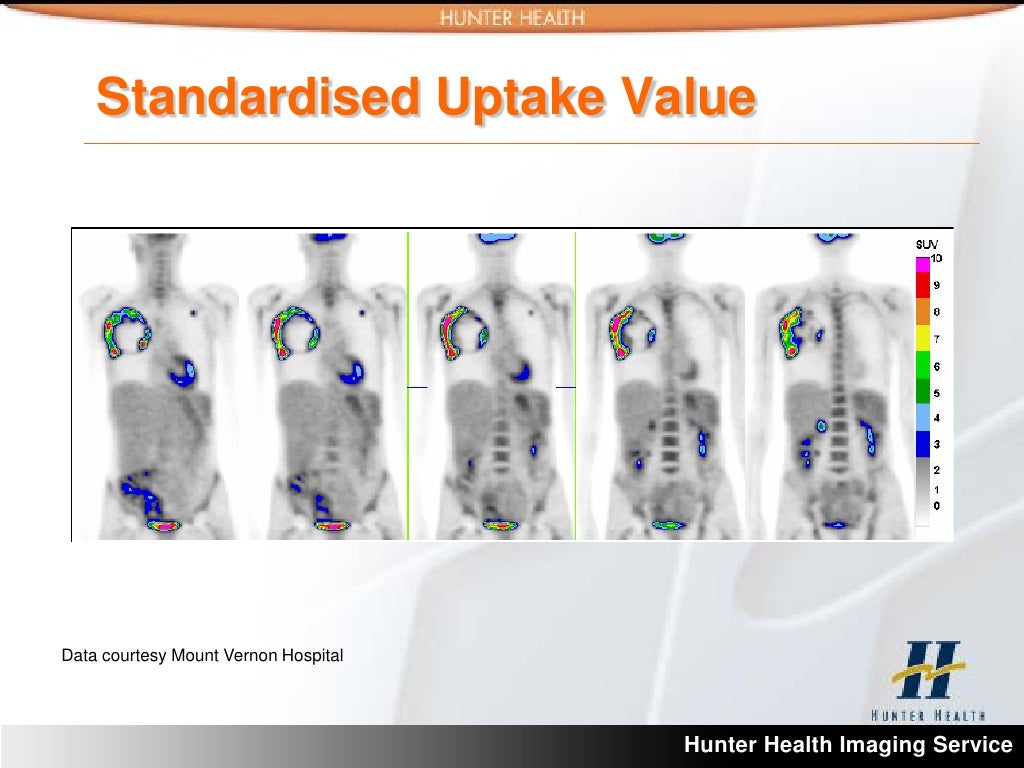
Abnormal SUV 5.8 PET Scan Image
An SUV 5.8 PET scan showing abnormal activity would present as a focal or regional area of significantly increased uptake. This increased uptake could indicate the presence of a tumor or inflammation. The abnormal region might display a distinct “hot spot” or a diffuse area of higher intensity compared to the surrounding healthy tissues. The degree of intensity difference would depend on the severity and type of pathology. Contrast between the abnormal region and the surrounding tissues would be significant, visually apparent.
Diagram of Tissue Metabolic Activity in SUV 5.8
The following diagram illustrates the metabolic activity of tissues in an SUV 5.8 PET scan, focusing on the uptake of radiotracers.
(Please note: A diagram cannot be directly inserted here. A diagram would visually represent different tissues (e.g., brain, muscle, tumor) with varying shades of color representing the SUV 5.8 values. Arrows could indicate the uptake of radiotracers by different tissues.)
This diagram would visually demonstrate the varying levels of metabolic activity across different tissues.
Radiotracer Types in SUV 5.8 PET Scans
Different radiotracers are used in SUV 5.8 PET scans, each targeting specific metabolic processes. These radiotracers allow for the detection and characterization of various pathologies.
- [18F]-FDG (Fluorodeoxyglucose): This is a common radiotracer used to detect glucose metabolism. It is particularly useful in identifying tumors that exhibit high glucose uptake, such as some cancers.
- [11C]-Choline: This radiotracer is used to assess the production of phospholipids, which are important for cell membranes. Increased uptake can indicate the presence of certain types of tumors, especially in the brain or prostate.
- Other Radiotracers: Other radiotracers are being developed to target specific metabolic pathways or cellular processes, enabling more refined analysis of metabolic activity. This allows for more precise characterization of different diseases.